Round to slightly oval nucleus with pink cytoplasm
• Grains
Raisinoid nuclei with pink cytoplasm
• Minimal to no cytologic atypia
Top Differential Diagnoses
• Darier disease
Histologically similar
Clinically different
– Multiple lesions in seborrheic distribution
• Acantholytic dyskeratotic acanthoma
Similar to WD
– Both have acantholysis and dyskeratosis
– Both are usually clinically solitary lesions
Unlike WD
– Not cup-shaped or associated with hair follicles
– Flat-based lesion
• Acantholytic squamous cell carcinoma
Infiltrative
Cytologic atypia
Atypical mitoses
Warty Dyskeratoma: Low-Power Architecture This is a low-power view of a warty dyskeratoma, a cup-shaped lesion. There is hyperkeratosis and parakeratosis above central acantholytic dyskeratosis with villi at the base .
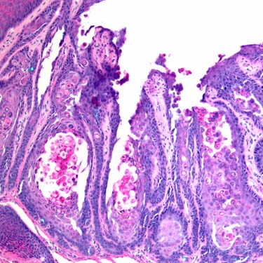
Warty Dyskeratoma: Acantholytic Dyskeratosis Acantholysis and dyskeratosis within the hyperplastic epithelial cup are seen here.
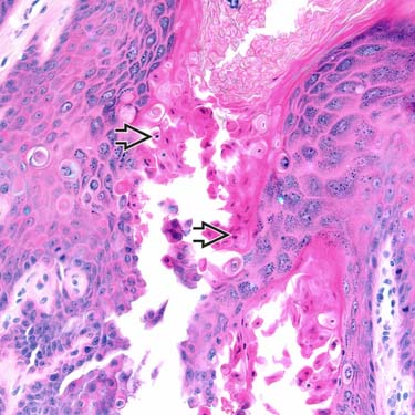
Acantholytic Dyskeratosis: Prominent Corp Ronds A high-power view shows the central acantholytic dyskeratosis in a warty dyskeratoma. There are corp ronds (central round to slightly oval shrunken nuclei with surrounding pink cytoplasm) .
TERMINOLOGY
Abbreviations
• Warty dyskeratoma (WD)
Definitions
• Cup-shaped epidermal proliferation
• Acantholysis is present
• Dyskeratosis (corp ronds and grains) is present
ETIOLOGY/PATHOGENESIS
Pathophysiology
• Germline mutations in ATP2A2 not described (as are seen in Darier disease)
• Human papilloma virus infection has not been associated with WD
CLINICAL ISSUES
Epidemiology
• Age
Middle-aged to older patients
Only gold members can continue reading. Log In or Register to continue


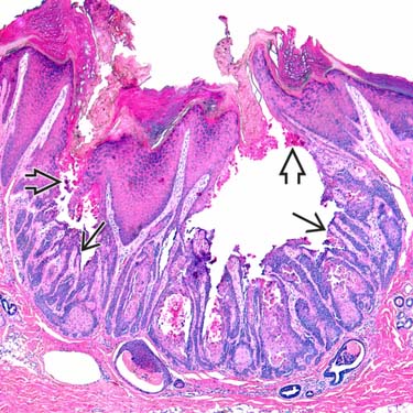
 with villi at the base
  .
.
 below hyperkeratosis.
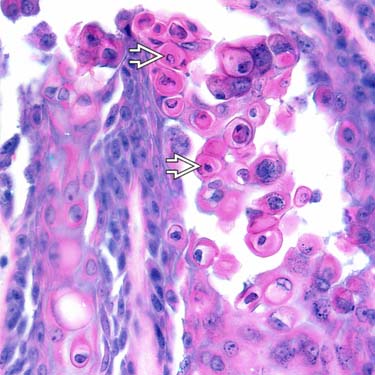
 .
.


