ITAN Clinical Clinically, an inverted type A nevus (ITAN) is compared to other pigmented papules. ITAN has an eccentrically placed dark papule upon an evenly pigmented flat area. The lesional borders are sharp in contrast to dysplastic nevus. (Courtesy R. McClintok, MD.)
ITAN at Scanning Magnification At scanning magnification, the nevus resembles an ordinary nevus with admixed melanophages .
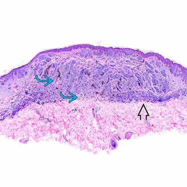
ITAN at Low Magnification At low power, the more epithelioid nests are identified in the middermis toward the bottom of the lesion . There is also a 2nd population of type B cells , resembling lymphocytes at this power.
ITAN at Intermediate Magnification Showing Maturation Under medium-power magnification, the type A nevus nests are contrasted to the type B cells . There is also a junctional component .
TERMINOLOGY
Abbreviations
• Inverted type A nevus (ITAN)
Synonyms
• Clonal nevus (superficial)
• Deep penetrating nevus (deep seated)
Definitions
• Benign melanocytic nevus with inverted maturation of dermal melanocytes
CLINICAL ISSUES
Presentation
• Recent color change in otherwise benign preexisting nevus
• Usually thin brown papule with eccentric small focus of dark pigmentation within less pigmented macule
Treatment
• Surgical approaches
Shave or simple excision is usually curative
Prognosis
• Excellent, no reported local recurrence or metastasis
IMAGING
General Features
• Dermoscopy shows regular and uniform brown globules with eccentric small blue-grey blotch
MICROSCOPIC
Histologic Features
• Collection of nests with distinctively epithelioid morphology, type A nevus cells
Cells located at or near bottom of lesion
Type A nevus cells are typically present in superficial dermis
– Location deep in reticular dermis gives its name, “inverted type A”
• May have variants or admixed with cell types
Purely type A (epithelioid nevus cells)
Type B (lymphoid nevus cells)
Type C (spindled nevus cells)
Dendritic blue cells
• Surrounded by numerous melanophages
Cytologic Features
• Nuclei of type A cells are round-to-plum/oval in shape with finely melanized cytoplasm
• Even chromatin with inconspicuous nucleoli
• Nuclear membrane is smooth
• Mitoses are absent to extremely rare
ANCILLARY TESTS
Histochemistry
• Examine additional deeper histological sections looking for mitoses
Only gold members can continue reading. Log In or Register to continue
 compared to other pigmented papules. ITAN has an eccentrically placed dark papule upon an evenly pigmented flat area. The lesional borders are sharp in contrast to dysplastic nevus. (Courtesy R. McClintok, MD.)
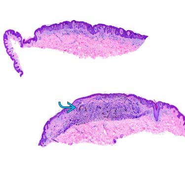
 .
.
 . There is also a 2nd population of type B cells
  , resembling lymphocytes at this power.
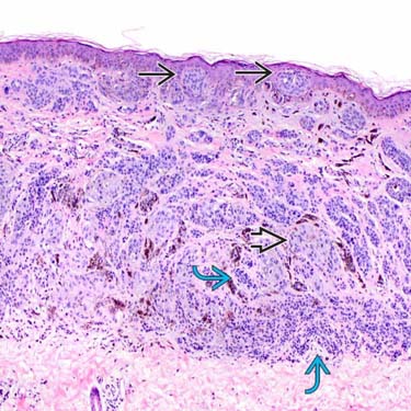
 are contrasted to the type B cells
  . There is also a junctional component
  .
.


