Spindle Cell/Pleomorphic Lipoma

 Variable admixture of bland spindle cells, ropey collagen, mature adipose tissue, myxoid stroma, and mast cells


• PL contains multinucleated tumor cells

• Morphologic variants: Fat-free, fat-poor, angiomatous


Ancillary Tests


• CD34(+); loss of nuclear retinoblastoma (Rb) expression

• Molecular: Deletion of RB1 (13q14)


Top Differential Diagnoses


• Atypical lipomatous tumor

• Neurofibroma

• Myxoid liposarcoma

• Solitary fibrous tumor

image
Spindle Cell Lipoma, Gross Specimen
Like conventional lipomas, spindle cell (SCL) and pleomorphic lipomas (PL) have a homogeneous yellow cut surface. Both tumors also often show subtle gray-white foci image 
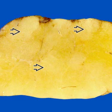
that are firm in consistency and correlate histologically with spindle cell areas containing ropey collagenous matrix.


image
Spindle Cell Lipoma at Low Magnification
At low power, SCL is composed of a mixture of adipose tissue and a variable myxoid to collagenous stroma containing bland spindle cells.

image
Ropey Collagen and Mast Cells
Thickened bundles and strips of collagen are a consistent finding in SCL/PL and a helpful diagnostic feature. Mast cells image 
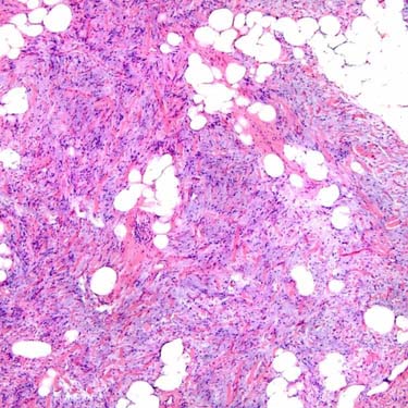
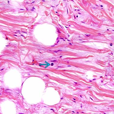
are also very commonly identified.
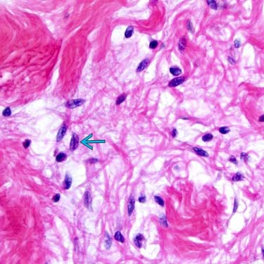
image
Bland Spindle Cells in Spindle Cell Lipoma
The lesional cells of SCL are cytologically bland and show bipolar or stellate cytoplasmic processes. Nuclear grooves may be identified in some cases image. Mitoses are rare.


TERMINOLOGY


Abbreviations


• Spindle cell lipoma (SCL)/pleomorphic lipoma (PL)


Definitions


• Benign adipocytic tumor with variable admixture of spindle cells, ropey collagen, and mature adipose tissue
image PL additionally contains multinucleated tumor giant cells

• SCL and PL are considered morphologic variations on same entity


CLINICAL ISSUES


Epidemiology


• Age
image Typically in older age group (> 50 years old)

image Very rare in patients < 20 years old

• Sex
image Strong male predilection
– Dermal SCL may be more common in females


Site


• Most occur superficially in subcutaneous tissue or dermis
image Exceptionally rare in deep soft tissues

• Shoulder, posterior neck, and back most common (80% of cases)
image Also face, oral cavity, trunk, extremities


Presentation


• Very similar to conventional lipoma
image Painless, slow-growing mass

image Often present for long duration

• Rarely arise in multiple sites (synchronous or metachronous)
image May be familial


Treatment


• Conservative surgical excision


Prognosis


• Benign
• Local recurrence is very rare

image Atypical findings in PL, such as lipoblasts and rare atypical mitoses, do not appear to affect behavior


MACROSCOPIC


General Features


• Well circumscribed
image Dermal tumors usually less circumscribed

• Yellow-tan cut surface

• Subtle gray-white foci correlate with spindle cell foci

• May show focal myxoid areas or be extensively gelatinous

• Very rare multinodular, plexiform growth pattern


Size


• Usually 3-5 cm (rarely > 10 cm)


MICROSCOPIC


Histologic Features


• SCL and PL exist as 2 ends of single morphologic spectrum
image Some tumors show morphology of SCL exclusively and some show morphology of PL exclusively

image Tumors demonstrating intermixed features or discrete areas of both SCL and PL are common

• SCL
image Variable admixture of 3 components
– Uniform, bland spindle cell proliferation
image Bipolar to stellate cytoplasmic projections

image May cluster together akin to schools of fish or show nuclear palisading

image May show nuclear grooves

image Mitotic figures rare to absent

– Mature adipocytes

– Bundles of ropey (thickened) collagen fibers

image Myxoid stroma is common and may be extensive

image Mast cells are very common and nearly constant finding

image Thick-walled vessels may show perivascular hyalinization or fibrosis or grow in focal ectatic staghorn pattern

• PL
image Characterized by variable numbers of multinucleated giant cells with dense eosinophilic cytoplasm and peripherally arranged nuclei (floret cells)
– Also cells with “smudgy” nuclei &/or nuclear pseudoinclusions

image Bland spindled cells, ropey collagen, mast cells, and adipose tissue also usually present

image May rarely contain multivacuolated lipoblasts

image Mitotic figures rare to absent (may be atypical)


Morphologic Variants


• Fat-free or low-fat SCL
image Minimal to no mature adipose tissue component

image May show abundant collagen, abundant myxoid stroma, or predominantly spindle cells

image Fat-free and low-fat forms of PL also exist

• Angiomatous SCL
image Also described as pseudoangiomatous SCL

image Contains prominent slit-like to ectatic vascular spaces lined by endothelial cells

Only gold members can continue reading. Log In or Register to continue

Stay updated, free articles. Join our Telegram channel

Apr 24, 2017 | Posted by in PATHOLOGY & LABORATORY MEDICINE | Comments Off on Spindle Cell/Pleomorphic Lipoma

Full access? Get Clinical Tree

Get Clinical Tree app for offline access