Typically show hyperchromatic-staining nuclei with small nucleoli
Cytoplasm with prominent melanin pigmentation
• Atypical Reed nevus
Often larger, more cellular lesions
Increased mitotic activity, especially in junctional component
Top Differential Diagnoses
• Spitz nevus
More common in children, on head and neck and trunk region (Reed nevi more often on extremities)
Composed of mixture of spindled and epithelioid-shaped melanocytes (more spindled cells in Reed nevi)
• Melanoma
More often occurs in older patients in sun-damaged skin
Greater cytologic atypia and pleomorphism
Dermal mitoses and lack of maturation with descent
Clinical Photograph of Reed Nevus Clinical photograph shows a pigmented spindle cell nevus on the knee of a young adult patient. This case shows dark pigmentation, except 1 area at the top of the lesion with depigmentation . (Courtesy P. Hsu, MD.)
Reed Nevus at Low Magnification Histologic examination of a Reed nevus shows fusion of rete ridges by a proliferation of junctional pigmented spindle cells, which show a characteristic streaming together pattern.
Reed Nevus With Junctional Nests Showing Bridging Higher magnification shows bridging across rete ridges by junctional nests of streaming pigmented spindle-shaped cells. The cells show hyperchromatic nuclei and heavily pigmented cytoplasm. Note the numerous darkly pigmented melanophages in the superficial dermis .
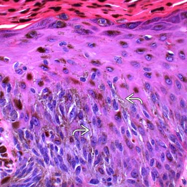
Reed Nevus at High Magnification High magnification of the intraepidermal component shows elongated spindle-shaped cells with pigmented cytoplasmic processes .
TERMINOLOGY
Abbreviations
• Pigmented spindle cell nevus (PSCN)
Synonyms
• Pigmented spindle cell nevus of Reed
Definitions
• Melanocytic proliferation, usually predominantly junctional, and composed of spindle-shaped cells with heavy cytoplasmic pigmentation
• Often considered variant of Spitz nevus, which occurs more frequently in adults
ETIOLOGY/PATHOGENESIS
Unknown
• May be related to solar exposure in some cases
CLINICAL ISSUES
Epidemiology
• Incidence
Relatively uncommon tumors
• Age
Typically young adults (< 40 years old)
• Sex
More common in females
• Ethnicity
Caucasian patients in most cases
Site
• Most often presents on extremities, especially leg
Classic presentation is on thigh of young woman
Presentation
• Pigmented lesion
Usually papular but can be nodular
Treatment
• Surgical approaches
Complete excision is curative
– Typically recommended in partially sampled lesions
– To allow for complete evaluation to exclude more atypical areas
– Also to prevent recurrence
Prognosis
• Excellent; may recur if incompletely excised but very low risk of developing melanoma
Only gold members can continue reading. Log In or Register to continue


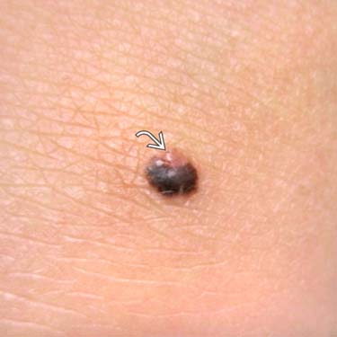
 . (Courtesy P. Hsu, MD.)
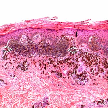
 pattern.
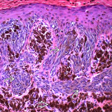
 of streaming pigmented spindle-shaped cells. The cells show hyperchromatic nuclei and heavily pigmented cytoplasm. Note the numerous darkly pigmented melanophages in the superficial dermis
  .
.
 .
.


