– Generally affects unilateral nipple/areola in older adults
Clear to pale cells with abundant cytoplasm scattered at all levels in epidermis
Nuclear atypia present
– Hyperchromatic nuclei
– Prominent eosinophilic cytoplasm
Variable mitotic activity
• Pagetoid dyskeratosis
Clusters of somewhat pale or clear keratinocytes
– Slightly shrunken nuclei
– Perinuclear halo
Possibly related to trauma
Cells do not mark with CK7
• Clear cell papulosis
Clinical presentation differs
– Child with multiple hypopigmented, white to flesh-colored papules on lower abdomen/groin area
Scattered, benign-appearing cells in basal layer of acanthotic epidermis
Cells stain with CK7
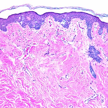
Toker Cell Hyperplasia Toker cell hyperplasia is a rare histopathologic finding. Toker cells, a normal component of the nipple epithelium, are increased in number and may mimic Paget disease of the nipple.
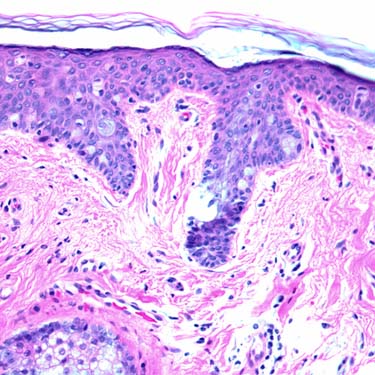
Toker Cell Hyperplasia at Higher Magnification At this magnification, the scattered, hyperplastic Toker cells in the epidermis mimic Paget disease of the nipple.
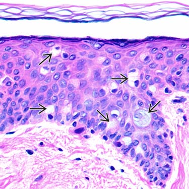
Toker Cell Hyperplasia at High Magnification This is a high-magnification view of Toker cell hyperplasia. Toker cells have bland nuclei with expanded, pale to clear-staining cytoplasm. They are in the basal and suprabasal layers of the epidermis.
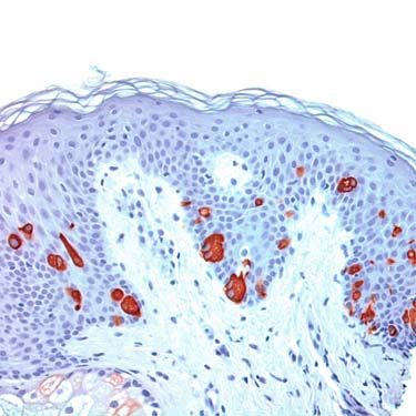
Toker Cell Hyperplasia: CK7 Immunohistochemistry In Toker cell hyperplasia, Toker cells are evident by light microscopy, and they are also highlighted by CK7 staining. The bland cells are scattered in the basal and suprabasal layers.
TERMINOLOGY
Definitions
• Increase in number of Toker cells in epidermis
• Absence of underlying ductal carcinoma
• Mimics Paget disease of nipple
• Awareness important to avoid overdiagnosis of Paget disease
CLINICAL ISSUES
Epidemiology
• Incidence
Toker cells present by light microscopy in ~ 10% of normal nipples
Toker cells also described in
Only gold members can continue reading. Log In or Register to continue


 have bland nuclei with expanded, pale to clear-staining cytoplasm. They are in the basal and suprabasal layers of the epidermis.