27
Suboccipital Region
The suboccipital region is in the upper part of the posterior aspect of the neck just below the occipital bone and deep to the trapezius, splenius capitis, and semispinalis capitis muscles. The suboccipital triangle contains the vertebral artery, the suboccipital nerve (C1), and the suboccipital venous plexus. Skeletal support for this region is provided by the occipital bone, the atlas (CI), and the axis (CII). These three bony structures form the atlanto-occipital and atlanto-axial joints. These articulations allow flexion, extension, and rotation of the head.
Muscles
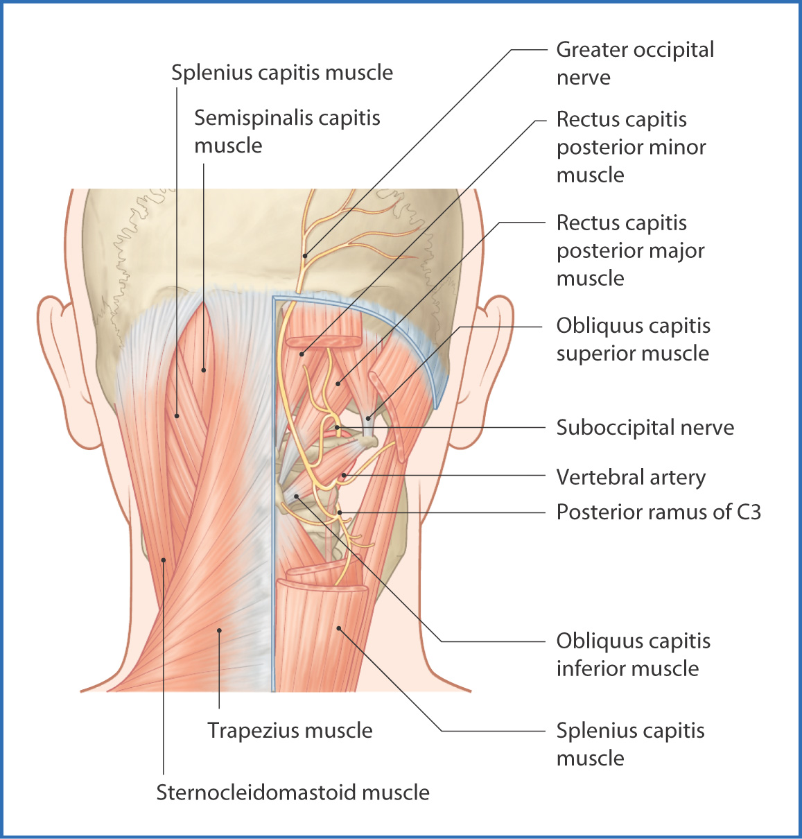
The four muscles of the suboccipital region lie deep to the semispinalis capitis muscle (Fig. 27.1 and Table 27.1). Three of these muscles form the borders of the suboccipital triangle:
- The inferolateral border is formed by the obliquus capitis inferior muscle.
- The superolateral border is formed by the obliquus capitis superior muscle.
- The superomedial border is formed by the rectus capitis posterior major muscle.
- The superolateral border is formed by the obliquus capitis superior muscle.

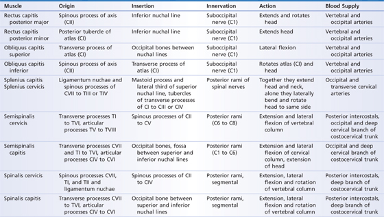
TABLE 27.1 Muscles of the Suboccipital Region

The fourth muscle, the rectus capitis posterior minor, does not take part in formation of the triangle, but it can be observed just medial to the rectus capitis posterior major muscle.
The main function of these muscles is to hold the head in the neutral position, but they also extend and rotate the head. They are innervated by branches of C1 (the suboccipital nerve).
Nerves
The posterior rami of the first four cervical nerves (C1 to C4) are in the suboccipital region (see Fig. 27.1). The suboccipital nerve (C1) enters the region by piercing the atlanto-occipital membrane and supplies all four suboccipital muscles. The greater occipital nerve (C2) passes inferiorly to the obliquus capitis inferior muscle and supplies sensation to the posterior skin of the scalp. The posterior rami of C3 and C4 supply the upper cervical muscles, scalp, and posterior skin of the neck.
Arteries
The structures in the suboccipital region receive blood from branches of the vertebral, occipital, and deep cervical arteries.
- The vertebral artery arises from the first part of the subclavian artery and ascends through the transverse foramina of the upper six cervical vertebrae on its way to the brain; its suboccipital part provides muscular branches to each of the suboccipital muscles.
- The occipital artery branches from the external carotid artery. In the suboccipital region, it lies on the obliquus capitis superior and semispinalis capitis muscles. Accompanied by the greater occipital nerve, the occipital artery pierces the trapezius muscle and ascends and divides into numerous branches to the posterior skin of the scalp.
- The deep cervical artery, a branch of the costocervical trunk, is located in the suboccipital region just deep to the semispinalis capitis muscle. Here, branches of the deep cervical artery anastomose with branches of the occipital artery.
- The occipital artery branches from the external carotid artery. In the suboccipital region, it lies on the obliquus capitis superior and semispinalis capitis muscles. Accompanied by the greater occipital nerve, the occipital artery pierces the trapezius muscle and ascends and divides into numerous branches to the posterior skin of the scalp.
Veins and Lymphatics
The deep cervical veins drain the suboccipital region and are deep to the semispinalis capitis muscle within the suboccipital triangle. They connect with the suboccipital venous plexus
Stay updated, free articles. Join our Telegram channel

Full access? Get Clinical Tree


