40
Anteromedial Thigh
The thigh is the part of the lower limb between the hip and knee joints. The muscles of the thigh are divided into anterior, medial, and posterior groups. In addition, the psoas major and iliacus muscles enter the thigh from the posterior abdominal wall and iliac fossa (Fig. 40.1). These two muscles combine to form the iliopsoas muscle. The tensor fasciae latae muscle, innervated by the superior gluteal nerve, is discussed here because of its close proximity to the anteromedial thigh muscles.
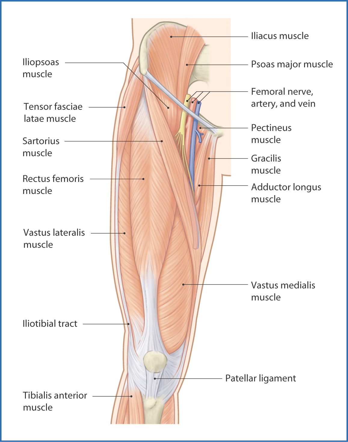
FIGURE 40.1 Muscles of the thigh—anterior view.
The femur is the largest bone in the body and the only osseous support for the thigh. It is long and slender with a rounded head that articulates with the acetabulum of the pelvis. Inferiorly, it has two large prominences—the medial and lateral condyles—that articulate with the tibia and patella at the knee joint.
Muscles
The muscles of the thigh are enclosed within a strong fascial sleeve—the fascia lata (Fig. 40.2). Three intermuscular septa arising from the fascia lata divide the thigh into three compartments: anterior, medial, and posterior compartments.
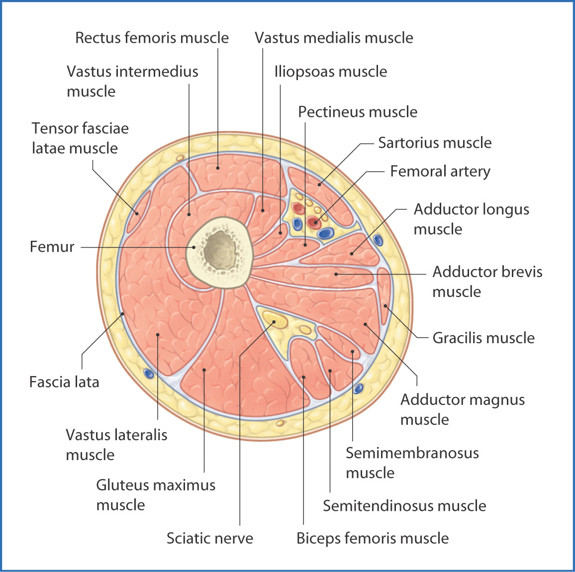
FIGURE 40.2 Cross section of the right thigh viewed from distal to proximal.
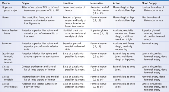
The muscles of the anterior compartment of the thigh (Table 40.1) are the sartorius and quadriceps femoris with its four heads: the rectus femoris, vastus medialis, vastus intermedius (which includes the articularis genus—a distinct muscle considered to be part of the vastus intermedius muscle that pulls the synovial membrane of the knee superiorly during knee joint extension), and vastus lateralis. As a group, the anterior thigh muscles (see Fig. 40.1) originate from either the anterior superior iliac spine of the pelvis or the shaft of the femur. The muscles insert onto the proximal end of the tibia through tendinous attachments, including the patellar tendon for the quadriceps femoris muscle and pes anserinus (“goose foot”), a three-pronged tendinous structure at the superior medial aspect of the tibia with attachment for the sartorius, gracilis, and semitendinosus muscles. The anterior thigh muscles flex the thigh at the hip joint and extend the leg at the knee.
TABLE 40.1 Anterior Thigh Muscles
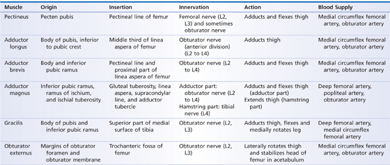
The muscles of the medial compartment of the thigh (Table 40.2) are the pectineus, adductor longus, adductor brevis, adductor magnus, gracilis, and obturator externus. The medial thigh muscles originate from the pubic rami and insert along the entire posterior length of the shaft of the femur (Fig. 40.3). Collectively, these muscles are the adductors of the thigh, with the exception of the obturator externus, which rotates the thigh laterally.
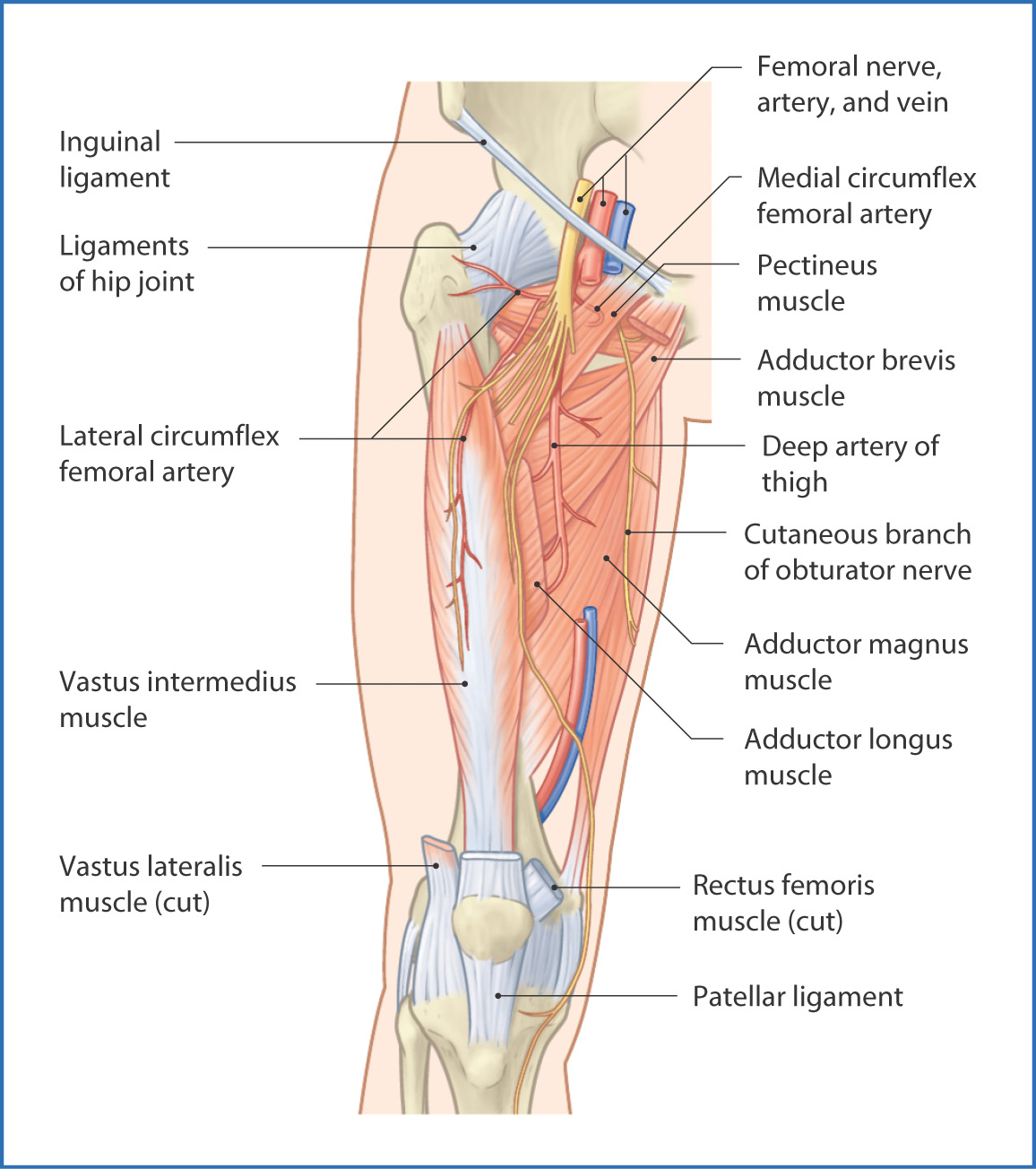
FIGURE 40.3 Deep muscles of the anterior part of the thigh.
TABLE 40.2 Medial Thigh Muscles
Two other muscles are related to the anteromedial part of the thigh:
- The tensor fasciae latae is lateral to the superior part of the vastus medialis muscle and tightens the iliotibial tract, which helps extend and stabilize the knee joint; it also aids in flexion and abduction of the hip joint.
- The iliopsoas muscle, formed from the union of the iliacus and psoas major muscles at the posterior abdominal wall, descends into the anteromedial part of the thigh beneath the inguinal ligament to insert onto the lesser trochanter of the femur and is the main flexor of the thigh at the hip joint.
The posterior thigh muscles are discussed in Chapter 42.
Nerves
The femoral nerve (L2 to L4) is the primary nerve to the anterior compartment of the thigh. Originating in the posterior abdominal wall as part of the lumbar plexus of nerves, the femoral nerve passes inferiorly and deep to the inguinal ligament in a groove formed by the psoas major and iliacus muscles to enter the anterior compartment of the thigh. From the femoral nerve, muscular branches run to the sartorius, pectineus, and each of the four heads of the quadriceps femoris. The terminal branch of the femoral nerve is the saphenous nerve, which enters the adductor canal and descends toward the medial compartment of the leg, where it conveys sensation from the skin of the medial arch of the foot and the medial part of the leg. Branches of the femoral nerve innervate the hip and knee joints.
The primary cutaneous nerves of the anteromedial part of the thigh are the anterior cutaneous branches of the femoral nerve (L2, L3). Several of these branches descend over the quadriceps femoris to supply the skin of the distal three quarters of the anterior surface of the thigh. The medial femoral cutaneous nerve supplies the skin of the distal two thirds of the medial surface of the thigh, and the lateral cutaneous nerve of the thigh (L2, L3), which is a direct branch of the lumbar plexus, supplies the skin over the tensor fasciae latae and vastus lateralis muscles in the lateral compartment of the thigh.
The obturator nerve (L2 to L4) enters the medial compartment of the thigh from the pelvis through the obturator canal. Superior to the adductor brevis muscle, the obturator nerve divides into anterior and posterior branches
Stay updated, free articles. Join our Telegram channel

Full access? Get Clinical Tree


