45
Posterior Leg
The posterior part of the leg (the calf) is between the knee and the ankle and contains superficial muscles that flex the ankle and deep muscles that flex the toes (Table 45.1). The bones of the leg, tibia, and fibula (see Chapter 44) provide support and muscle attachment for the posterior compartment of the leg.
TABLE 45.1 Posterior Leg Muscles
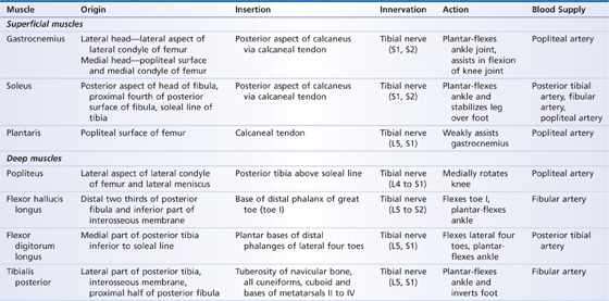
Muscles
The muscles of the posterior compartment of the leg are divided into superficial and deep groups by a thickened layer of deep fascia. The three superficial muscles plantar-flex the ankle and are the gastrocnemius, soleus, and plantaris muscles (Fig. 45.1). They have a general origin from the femoral condyles and the superior part of the tibia and fibula. All three muscle tendons fuse inferiorly to the calcaneus through the large calcaneal tendon (Achilles’ tendon).
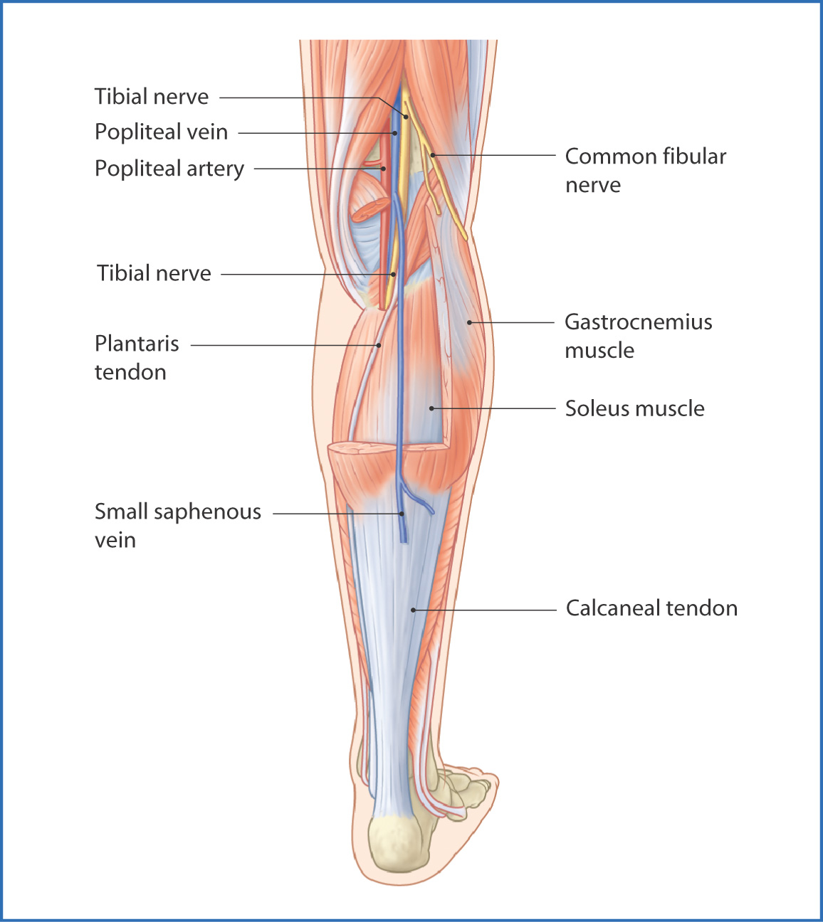
FIGURE 45.1 Superficial muscles of the posterior leg.
The four deep muscles in the posterior compartment of the leg are the popliteus, flexor hallucis longus, flexor digitorum longus, and tibialis posterior muscles (Fig. 45.2). With the exception of the popliteus muscle, they have a general origin from the posterior aspect of the tibia and fibula and plantar-flex the foot and toes. The popliteus muscle originates from the lateral condyle of the femur, inserts onto the posterior aspect of the superior surface of the tibia, and medially rotates the leg, thus unlocking and flexing the hyperextended knee.
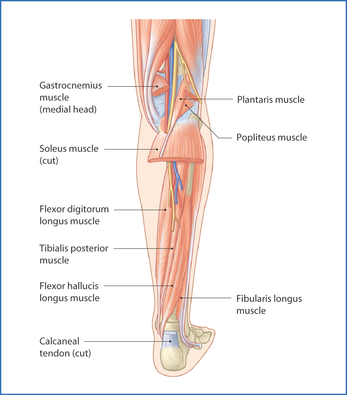
FIGURE 45.2 Deep muscles of the posterior leg.
Nerves
The tibial nerve (L4 to S3) originates in the thigh as a division of the sciatic nerve and descends into the leg through the popliteal fossa. At this point it gives off the medial sural cutaneous nerve, which unites with the lateral sural cutaneous nerve (from the fibular nerve) to form the sural nerve (see Fig. 45.6). The sural nerve conveys sensation from the skin of the posterolateral surface of the leg and foot.
From the popliteal fossa, the tibial nerve enters the posterior compartment of the leg and innervates all superficial and deep posterior leg muscles. It continues distally and is located between the flexor digitorum longus and flexor hallucis longus tendons at the level of the medial malleolus of the ankle. On entering the foot, it divides into the medial and lateral plantar nerves.
Arteries
Branches of the popliteal artery supply the posterior compartment of the leg.
The popliteal artery descends through the popliteal fossa of the knee and at the distal border of the popliteus muscle gives off the anterior tibial artery, which travels forward between the leg bones to the anterior compartment of the leg. Before the anterior tibial artery pierces the interosseous membrane, it usually gives off the posterior tibial recurrent artery medially and a small circumflex fibular artery laterally. Both these branches anastomose with the inferior lateral genicular artery.
The posterior branch of the popliteal artery—the posterior tibial artery—gives off muscular branches to the soleus and flexor digitorum longus muscles. Its largest branch is the fibular artery
Stay updated, free articles. Join our Telegram channel

Full access? Get Clinical Tree


