26
Vertebral Column
The vertebral column is a series of bones (vertebrae) stacked on top of each other to support the weight of the body. It is a central attachment point for muscles and bones of the appendicular skeleton. There are 33 vertebrae: 7 cervical, 12 thoracic, 5 lumbar, 5 fused sacral, and 4 fused coccygeal vertebrae.
Typically, a vertebra has a vertebral body, two pedicles, two laminae, two transverse processes, and a single spinous process. The vertebral foramen is the space between the vertebral body, pedicles, and laminae. Consecutive foramina create the vertebral canal, which contains the spinal cord and meninges. Most vertebrae possess a pair of superior and inferior articular processes (Fig. 26.1). The facet joints between these processes confer an interlocking arrangement that limits anterior and posterior dislocation of the spinal column.
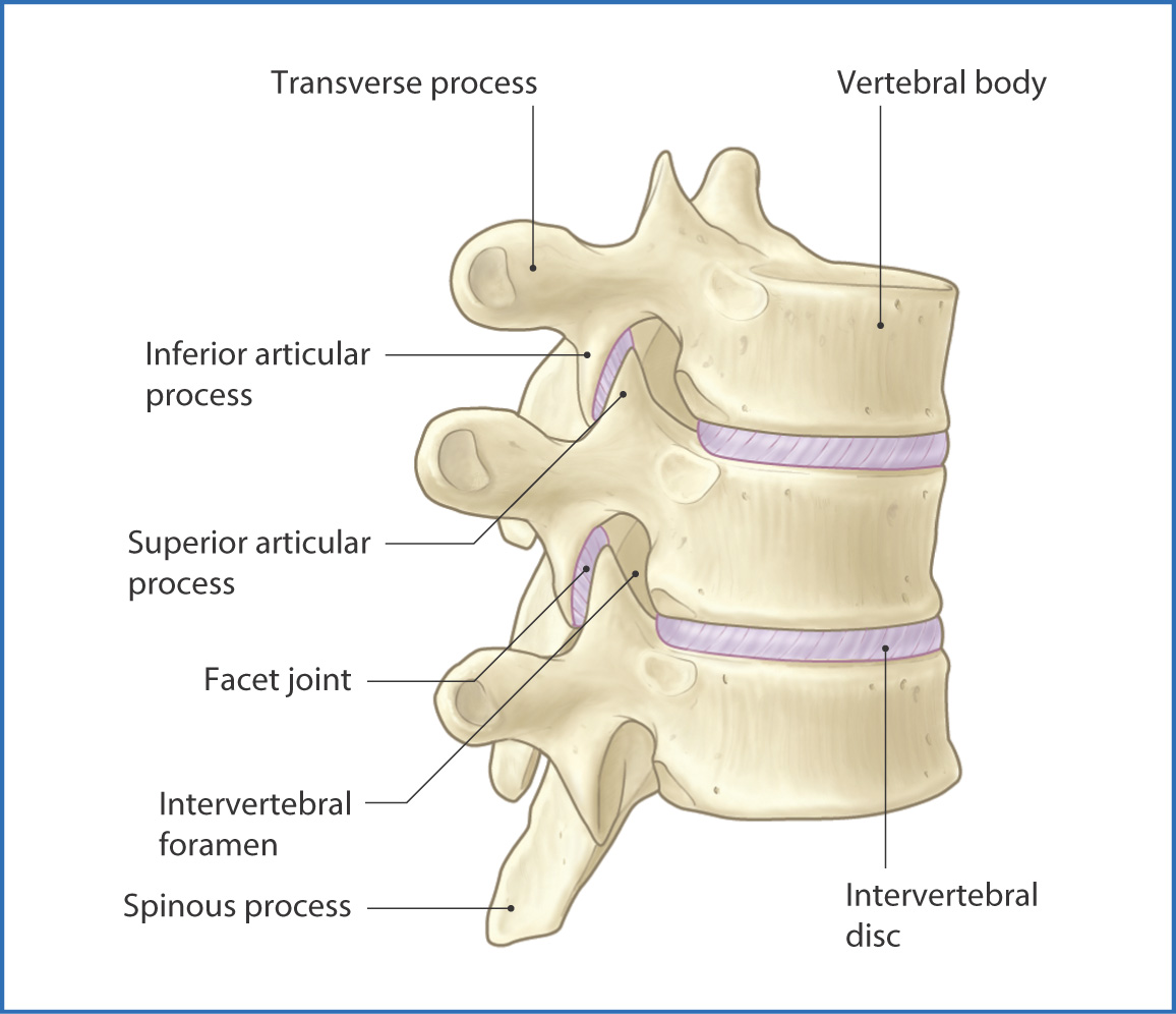
FIGURE 26.1 Anterolateral view of the vertebral column.
The vertebral bodies are united by fibrocartilaginous intervertebral discs. The outer part of a disc is the tough anulus fibrosus (ring) and the soft inner part is the nucleus pulposus. The combination of these two parts is such that the intervertebral disc can act as a shock absorber for the vertebral column and provide some flexibility.
At birth the vertebral column is concave anteriorly in the thoracic and sacral regions. These are the primary curves of the spine. The secondary (convex) curves of the cervical and lumbar regions develop after birth with elevation and extension of the infant’s head and when the child assumes an erect position when learning to walk. The joints, ligaments, and muscles of the back (see Chapter 28) stabilize and facilitate movement of the vertebral column. Several ligaments provide support to the vertebral column (Fig. 26.2, Table 26.1), including
- the anterior longitudinal and posterior longitudinal ligaments on the anterior and posterior surfaces of the vertebral body, which together with the intervertebral discs make up the joints between vertebral bodies
- the ligamentum flavum between adjacent laminae
- the supraspinous ligaments, which connect the tips of the spinous processes
- the interspinous ligaments between the spinous processes
- the ligamentum flavum between adjacent laminae
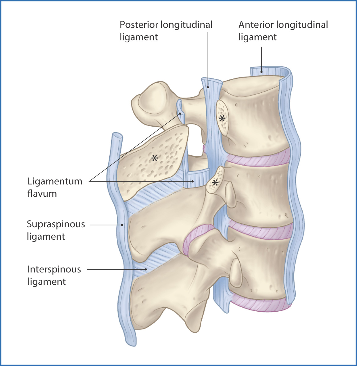
FIGURE 26.2 Ligaments of the spine. Asterisks indicate cut surfaces.
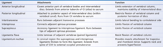
TABLE 26.1 Ligaments of the Vertebral Column
Vertebral Types
The cervical vertebrae are distinguished from other vertebrae by their small vertebral bodies, bifid spinous processes (except CVII), and transverse foramina within the transverse processes (which allow passage of the vertebral artery).
The atlas (CI) has no body or spinous process, but it has two lateral masses connected by the anterior and posterior arches. It articulates with the occipital bone of the skull through the atlanto–occipital joint, which enables flexion, extension, and lateral bending (flexion) of the head. The axis (CII) has a unique finger-like projection—the dens (odontoid process)—that articulates with the anterior arch of the atlas (CI). This unique articulation allows rotation of the head. There are also two joints between the lateral masses of the atlas and axis, the atlanto-axial joints.
Vertebra CVII has a large palpable spinous process and is known as the vertebra prominens.
The thoracic vertebrae are characterized by articular facets on their vertebral bodies and transverse processes for articulation with the ribs (except TXI and TXII) at the costovertebral joints. Their broad laminae and downward-projecting spinous processes overlap with those of the vertebra beneath and increase rigidity in this region.
Each lumbar vertebra (Fig. 26.3) has a large vertebral body to withstand the accumulated weight of the body. The superior articular processes (the mammillary process) are enlarged.
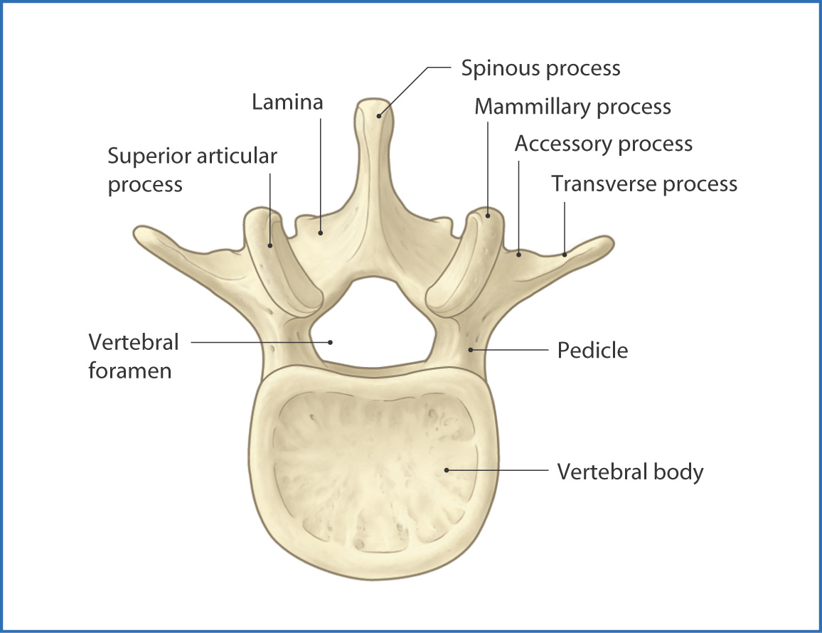
FIGURE 26.3 Superior view of a typical lumbar vertebra.
The sacrum, a fusion of five vertebrae, is a triangular-shaped bone that forms the concave posterior wall of the pelvis. It is weight bearing and has foramina for nerves from the lumbosacral plexus (see Chapter 35). At the inferior end of the vertebral column is the small coccyx (tail bone), which is made up of four fused vertebrae. It serves as an attachment point for pelvic ligaments and muscles.
Nerves
Innervation of the vertebrae, ligaments, joints, muscles, and skin of the vertebral column is from the posterior rami of spinal nerves as they exit from their respective intervertebral foramina (Fig. 26.4).
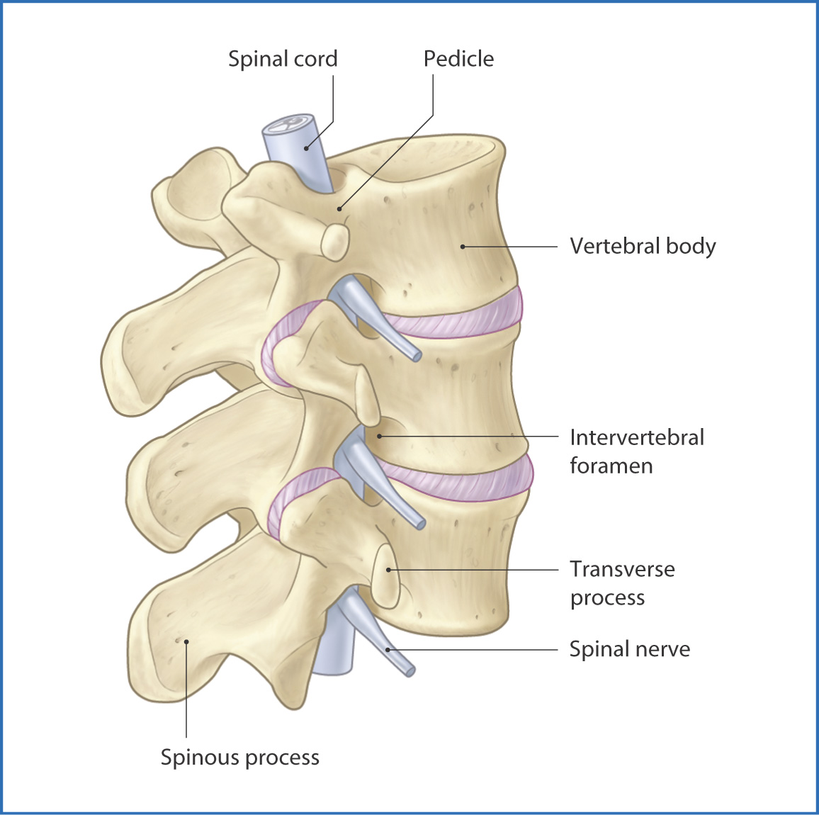
FIGURE 26.4 Posterolateral view of the vertebral column.
Arteries
Blood is supplied to the vertebrae by segmental spinal branches of the ascending cervical, vertebral, intercostal, lumbar, and lateral sacral arteries.
Veins and Lymphatics
Venous drainage of the external vertebral column is provided by a network of valveless veins that envelop each vertebra. These veins connect superiorly with the venous dural sinuses from the skull and brain and inferiorly with the pelvic veins. In the thorax, the external vertebral venous plexus connects with the azygos vein and the superior and inferior vena cavae.
Stay updated, free articles. Join our Telegram channel

Full access? Get Clinical Tree


