41
Hip Joint
The hip joint is a ball-and-socket synovial joint that connects the lower limb to the pelvis (Fig. 41.1). It is formed by the head of the femur and the acetabular fossa of the hip bone and is a multi-axial joint that allows multiple movements (flexion, extension, abduction, adduction, lateral and medial rotation, and circumduction). The hip joint is supported by muscles and ligaments in the hip area (Table 41.1).
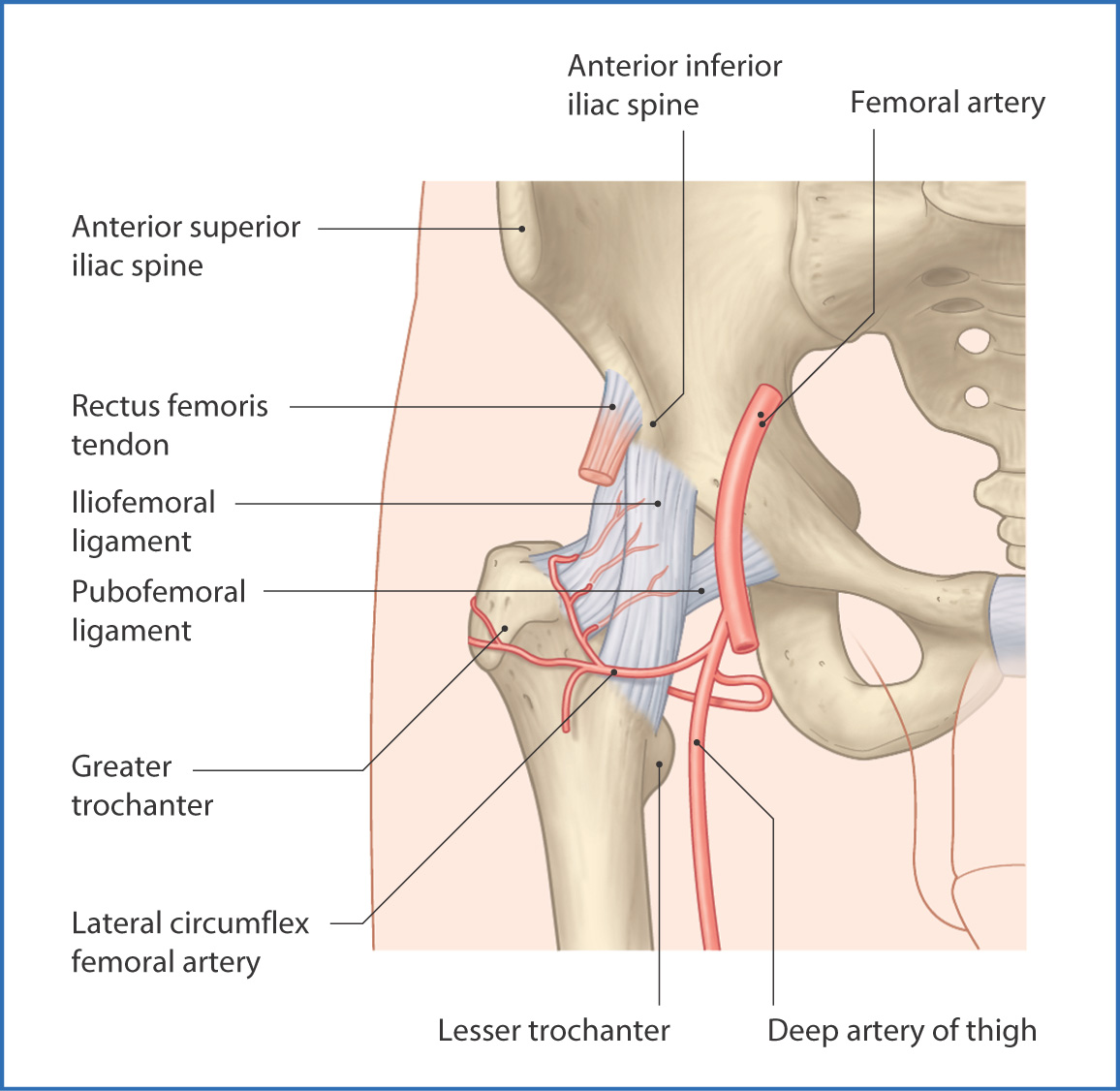
FIGURE 41.1 Hip joint (anterior view).
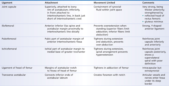
TABLE 41.1 Ligaments of the Hip Joint
The acetabulum is a cup-shaped fossa on the lateral aspect of the pelvis. Parts of the ilium, ischium, and pubic bones contribute to it. The acetabular labrum is a fibrocartilage lip that attaches to the edges of the acetabulum. This provides a deeper socket for the head of the femur and helps prevent easy dislocation of the hip. The anterior rim of the acetabular labrum is partly deficient at the acetabular notch and is bridged by the transverse acetabular ligament.
The femoral head has a central depression (fovea for the ligament of the head) for attachment of the vascular intracapsular ligament of the head of the femur. The artery to the head of the femur and accompanying veins and nerves run with the intracapsular ligament and nourish and innervate the head of the femur.
Surrounding the hip joint is a very strong joint capsule. It completely encloses the hip joint but is thin anteriorly at the location of the tendon of the iliopsoas muscle. Proximally, the joint capsule is attached to the acetabular labrum and the edge of the acetabular notch. Distally, it is attached to the femur along the intertrochanteric line and to the neck of the femur approximately 1 cm superior to the intertrochanteric crest.
The fibrous joint capsule is reinforced anteriorly by the strong Y-shaped iliofemoral ligament, which joins the anterior superior iliac spine and acetabular margin of the pelvis proximally to the intertrochanteric line of the femur distally. The iliofemoral ligament tightens when standing and contributes to maintenance of posture and limitation of joint hyperextension.
The pubofemoral ligament is on the inferior and anterior surfaces of the joint. It originates from the inferior half of the acetabular margin and inserts laterally onto the inferior fibers of the iliofemoral ligament. The pubofemoral ligament tightens during extension and abduction, thus preventing over-abduction of the joint.
The ischiofemoral ligament, the thinnest of the three hip joint ligaments, is on the posterior surface of the joint capsule (Fig. 41.2). It originates on the inferior part of the acetabulum and inserts laterally onto the superior surface of the neck of the femur just medial to the greater trochanter. The ischiofemoral ligament helps prevent hyperextension of the hip joint.
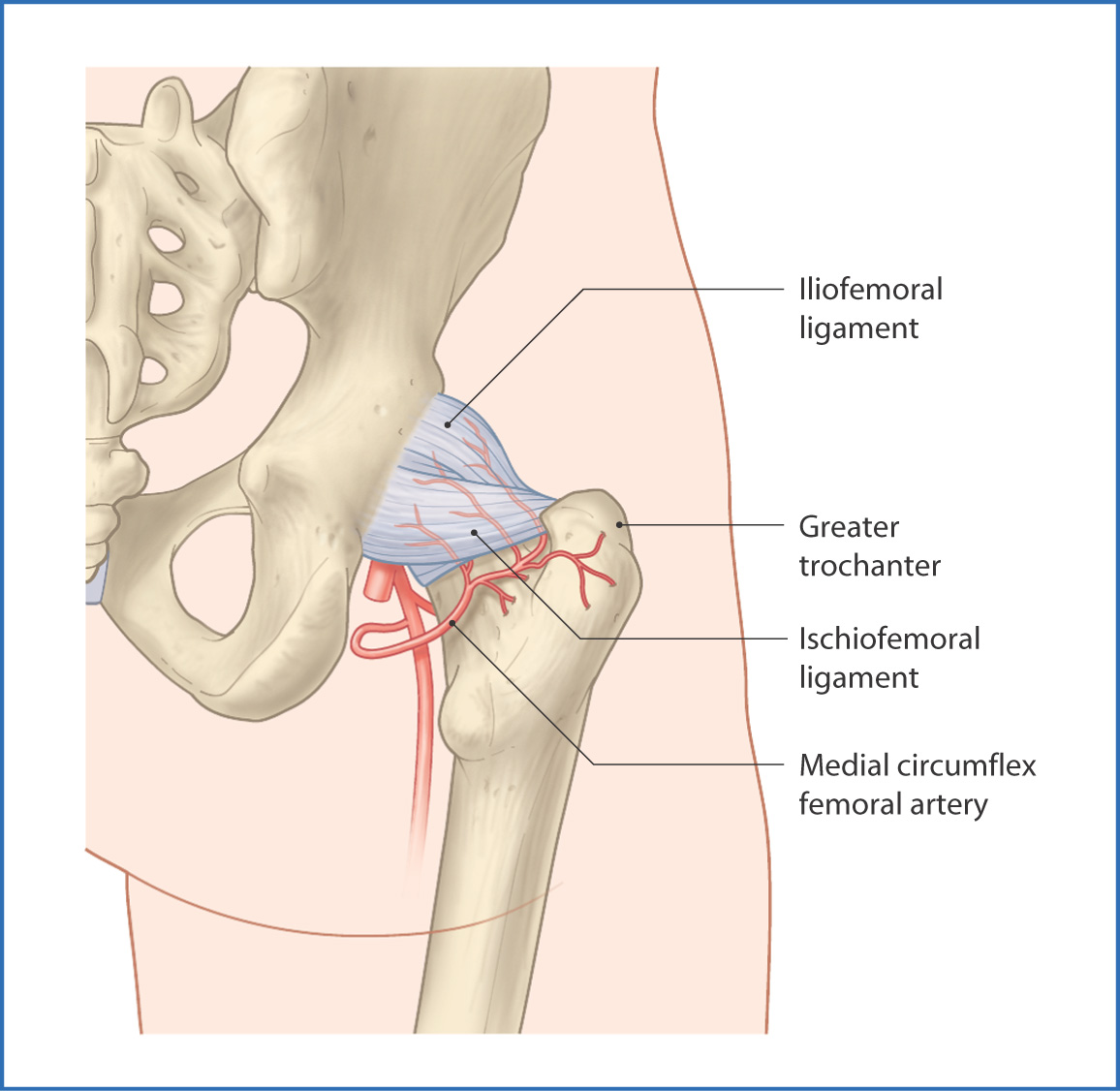
FIGURE 41.2 Main ligaments of the hip joint (posterior view).
Muscles
The muscles of the hip and thigh traverse the hip joint and provide support. From the anterior side, the anteromedial thigh muscles pass from the pelvis to the femur and move and support the joint (see Chapter 40). Posteriorly, the hip joint is supported by the muscles of the gluteal region and posterior compartment of the thigh (see Chapter 42).
Nerves
Innervation of the hip joint is from small branches of the nerves that pass nearby. The femoral nerve (L2 to L4) innervates the hip joint as it passes over it anteriorly (see Figs 40.1 and 40.3). The posterior side of the hip joint is innervated by the nerve to the quadratus femoris
Stay updated, free articles. Join our Telegram channel

Full access? Get Clinical Tree


