19
Arm
The arm is the part of the upper limb that extends from the shoulder to the elbow. The upper part of the arm receives tendons from the shoulder and scapular region, whereas the lower part sends tendons to the forearm. This unique arrangement enables the arm to take part in
- flexion, extension, abduction, adduction, and circumduction at the glenohumeral joint
- flexion and extension at the elbow joint
The arm is supported by the humerus (arm bone), the upper end of which (the head of the humerus) is ball shaped to enable articulation with the glenoid cavity of the scapula. The anatomical neck separates the head from the proximal part of the shaft of the humerus and has two prominences:
These tubercles are separated by the intertubercular sulcus, which receives the tendon of the long head of the biceps brachii muscle. The region where the tubercles join the shaft of the humerus is the surgical neck of the humerus and is a common fracture site. The long slender shaft of the humerus provides attachment areas for the arm muscles. Near its distal end, the humerus is expanded medially and laterally by the medial and lateral supracondylar ridges and the subjacent medial and lateral epicondyles. The distal end of the humerus has articular surfaces:
- The trochlea articulates with the ulna.
- The capitulum articulates with the radius.
- The olecranon fossa on the posterior surface accommodates the olecranon of the ulna at the elbow joint during extension.
- The capitulum articulates with the radius.
Muscles
The deep fascia that surrounds the arm gives rise to the medial and lateral intermuscular septa of the arm. These fascial walls divide the arm into anterior (flexor) and posterior (extensor) compartments:
- The medial intermuscular septum of the arm extends from the medial lip of the intertubercular sulcus at the superior aspect of the humerus to the inferiorly located medial epicondyle.
- The lateral intermuscular septum of the arm extends from the lateral lip of the intertubercular sulcus at the superior aspect of the humerus to the inferiorly placed lateral epicondyle.
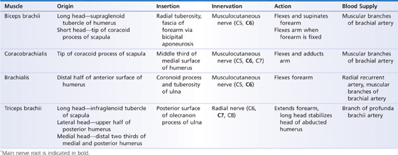
Superiorly, compartmentalization of the arm is completed by blending of the medial and lateral intermuscular septa with the deep fascia of the coracobrachialis and deltoid muscles, respectively. The posterior compartment of the arm contains the triceps brachii muscle, which has three heads originating superiorly from the humerus and scapula and joining inferiorly to insert onto the olecranon process of the ulna (Figs. 19.1 and 19.2). This arrangement makes the triceps brachii a strong extensor of the forearm at the elbow joint. The radial nerve (from the brachial plexus) provides innervation, and the profunda brachii (deep brachial artery branching off the brachial artery) supplies blood. The ulnar nerve passes through the inferior part of the posterior compartment of the arm on its way to the hand.
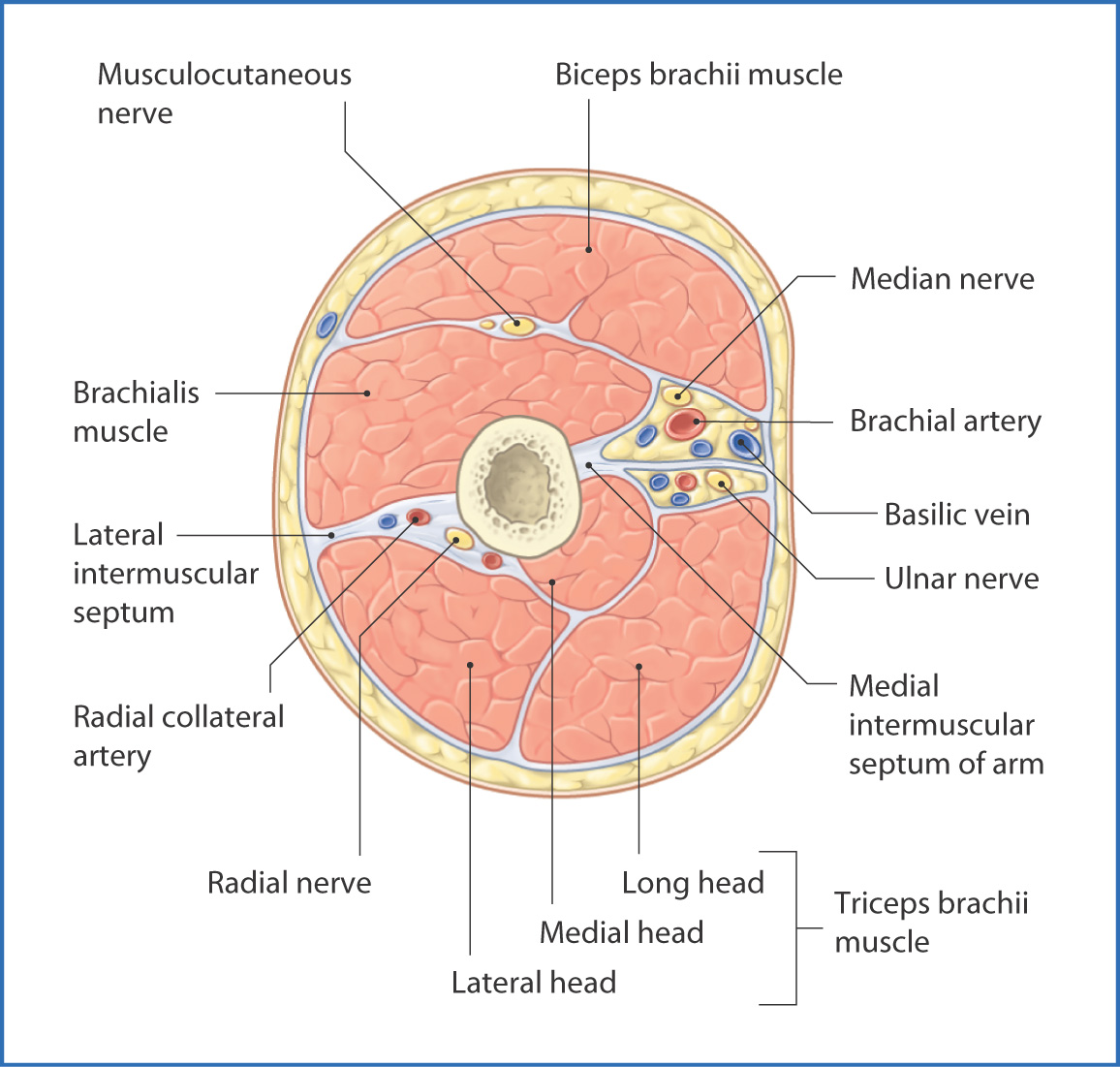
FIGURE 19.1 Cross section through the mid right arm (viewed from distal to proximal).
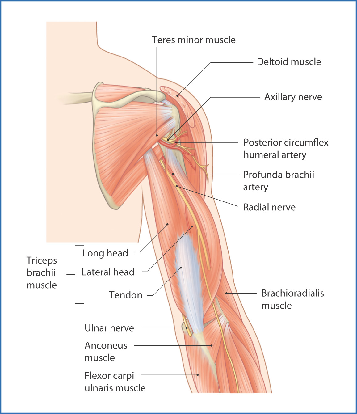
FIGURE 19.2 Arm (posterior view).
The flexor compartment of the arm contains the biceps brachii muscle, which has two heads of origin, and the coracobrachialis and brachialis muscles (Fig. 19.3). These three muscles have points of origin on either the scapula or the humerus, or on both, and insert inferiorly onto the humerus and the proximal ends of the radius and ulna. This arrangement enables the anterior arm muscles to help flex the arm at the glenohumeral joint and to flex the forearm at the elbow joint. Innervation of the anterior arm muscles is provided by the musculocutaneous nerve (from the brachial plexus), and blood is supplied by the brachial artery (Table 19.1).
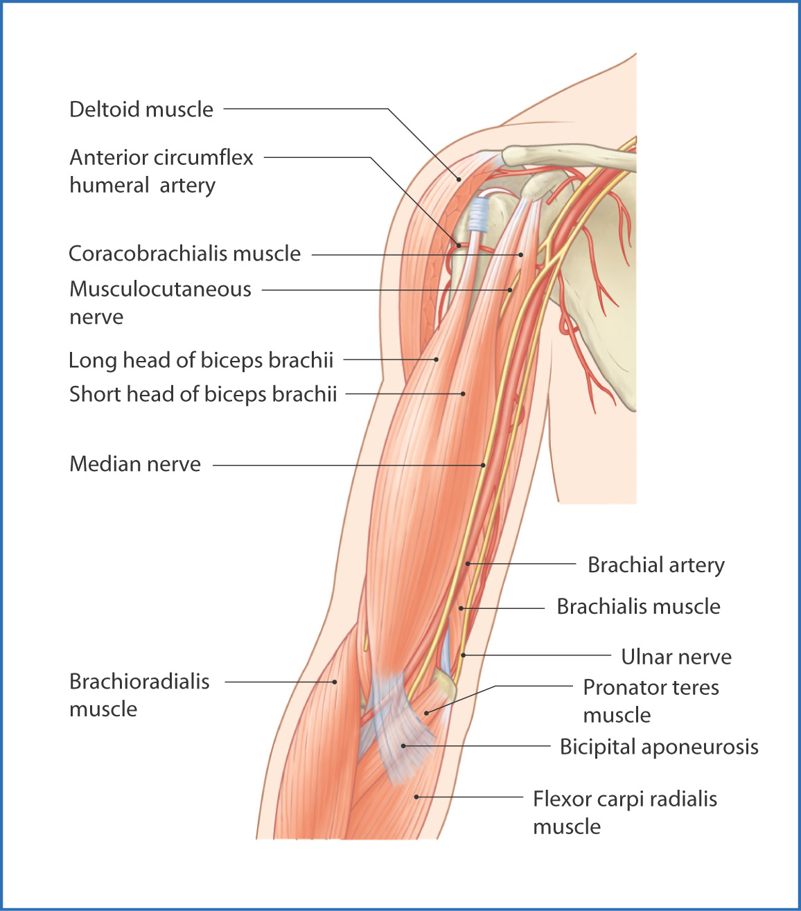
FIGURE 19.3 Arm (anterior view).
TABLE 19.1 Arm Muscles*
Nerves
The anterior and posterior compartments of the arm are innervated by the musculocutaneous and radial nerves. In addition, two major nerves from the brachial plexus (the median and ulnar nerves) pass through the arm to innervate structures in the forearm and hand.
The musculocutaneous nerve (C5 to C7 from the lateral cord of the brachial plexus) pierces the coracobrachialis muscle and descends between the biceps brachii and brachialis muscles to the lateral aspect of the arm. It supplies the three flexor muscles of the arm, sends an articular branch to the elbow joint, and terminates as the lateral cutaneous nerve of the forearm, which conveys sensation from the lateral surface of the forearm.
The radial nerve
Stay updated, free articles. Join our Telegram channel

Full access? Get Clinical Tree


