11
Submandibular Region
The submandibular region lies between the mandible and hyoid bones and is formed by the submental and submandibular triangles. It contains the submandibular gland. Skeletal support is provided by the mandible and the hyoid bone. The mandible is the site of attachment for some of the suprahyoid muscles and all muscles of mastication. In adults the mandible, which normally contains 16 permanent teeth, articulates with the temporal bone of the skull through the temporomandibular joint.
The hyoid bone is at the level of vertebra CIII, does not articulate with other bones, and is suspended by ligaments and the suprahyoid and infrahyoid muscles. It is linked to the thyroid cartilage through the thyrohyoid membrane. The hyoid muscles therefore move the larynx both superiorly and inferiorly during swallowing and speech.
Muscles
The most superficial muscle of the submandibular region is the digastric muscle (Table 11.1), which has two bellies (anterior and posterior) connected by an intermediate tendon attached to the hyoid bone via a “fascial pulley” (Fig. 11.1). The stylohyoid muscle extends from the styloid process of the temporal bone to the hyoid bone. The stylohyoid muscle is perforated by the tendon of the digastric muscle at the hyoid bone to create the fascial pulley.
TABLE 11.1 Muscles of the Submandibular Region (Suprahyoid Muscles)
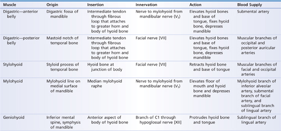
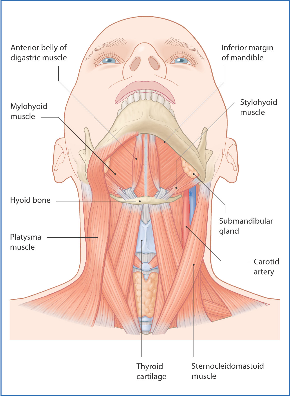
FIGURE 11.1 Submandibular region and its major muscles.
The mylohyoid muscle is deep to the anterior belly and superior to the posterior belly of the digastric muscle and forms the floor of the mouth; it is a sheet-like muscle that extends from the mylohyoid line of the mandible to the hyoid bone. Lymph nodes and neurovascular structures of the submandibular region are superficial to the mylohyoid muscle.
In the midline and deep to the mylohyoid muscle, the geniohyoid muscle emerges from the internal surface of the mandible and attaches to the hyoid bone (Fig. 11.2). Just lateral and posterior to the geniohyoid, the hyoglossus muscle attaches the hyoid bone to the base of the tongue.
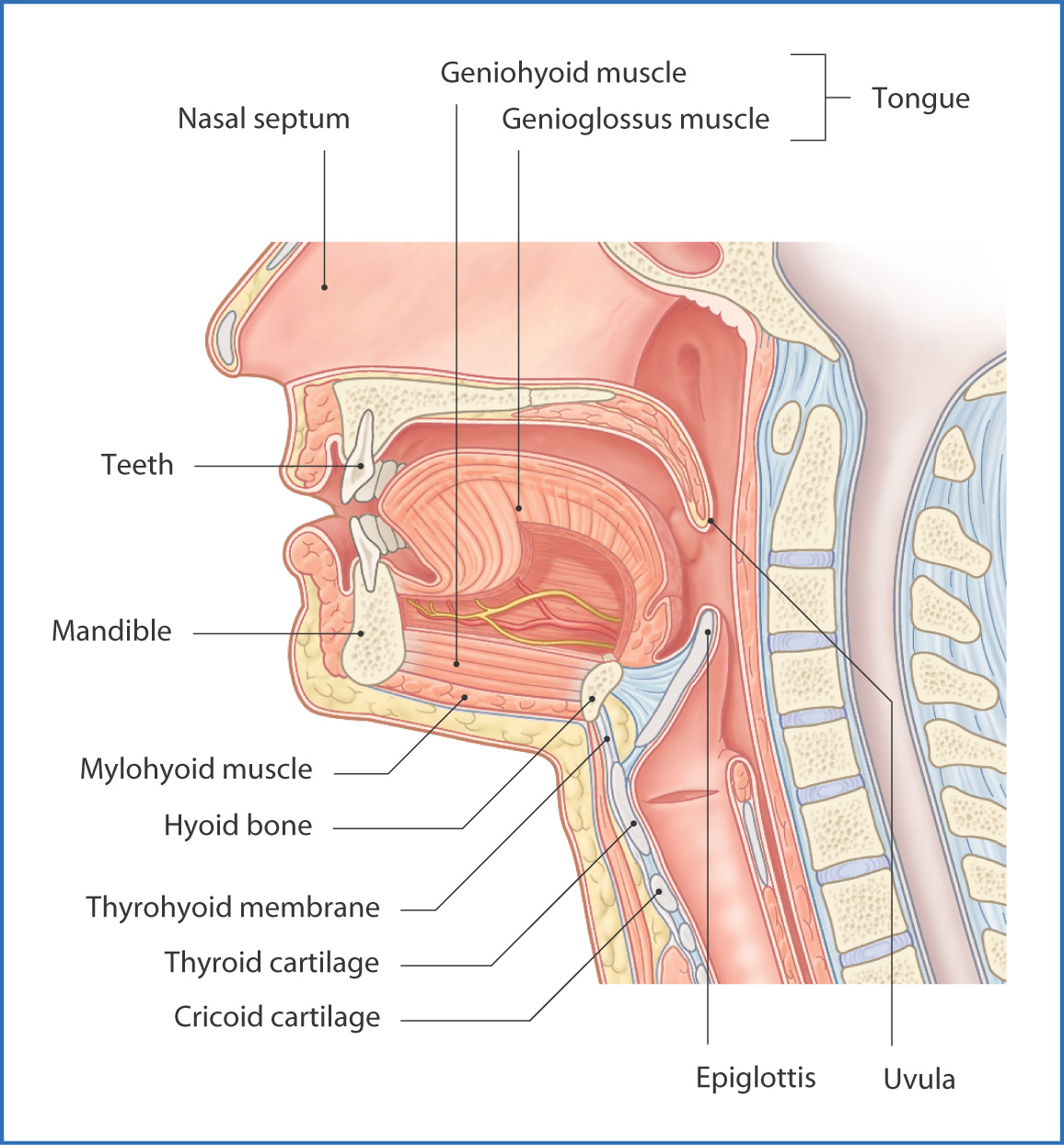
FIGURE 11.2 Major structures of the submandibular region in a hemisected specimen.
Nerves
Several cranial nerves and their branches are associated with the submandibular region. The mandibular nerve [V3] division of the trigeminal nerve [V] gives off two nerves to this area:
- The first branch—the nerve to mylohyoid (from the inferior alveolar nerve)—passes along the mylohyoid groove on the medial surface of the mandible and innervates the mylohyoid muscle and the anterior belly of the digastric muscle.
- The second branch—the lingual nerve—passes anteriorly and inferiorly from its origin in the infratemporal fossa toward the tongue; carries general sensation from the anterior two thirds of the tongue, the mucosa of the floor of the mouth, and the mandibular lingual gingiva; and also contains taste and preganglionic parasympathetic nerve fibers from the facial nerve [VII] via the chorda tympani nerve.
Stay updated, free articles. Join our Telegram channel

Full access? Get Clinical Tree


