Low magnification shows a noncircumscribed lesion in the dermis and subcutis composed of an admixture of thin-walled dilated blood vessels and sheets of spindle cells.
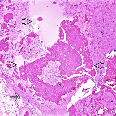
Low magnification of a spindle cell hemangioma shows a well-circumscribed intravascular tumor with ectatic vascular spaces
 and a cellular spindle cell proliferation. There is a remnant of a preexisting vessel at the periphery
  .
.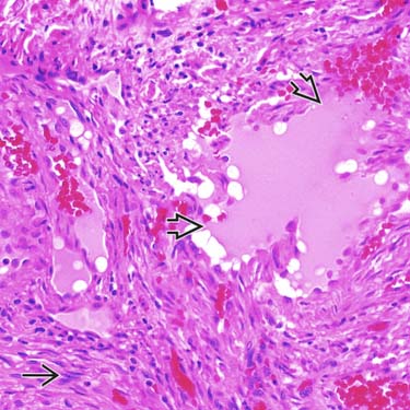
High magnification of spindle cell hemangioma shows straight/curved fascicles of uniform spindle cells
 with irregularly shaped vascular spaces
  and small areas of hemorrhage.
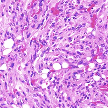
High magnification shows the spindled tumor cells and the characteristic vacuolated endothelial cells (so-called blister cells).
CLINICAL ISSUES
Epidemiology


Treatment
• Surgical approaches


Stay updated, free articles. Join our Telegram channel
Full access? Get Clinical Tree
