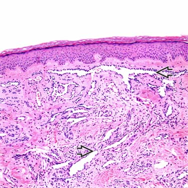
Retiform Hemangioendothelioma at Low Magnification Low-magnification examination of a retiform hemangioendothelioma (RHE) shows superficial dermal involvement by irregular, elongated, branching vascular spaces .
Retiform Hemangioendothelioma at Higher Magnification Higher magnification view shows a proliferation of elongated vessels lined by hyperchromatic-staining hobnailed endothelial cells . The surrounding stroma shows fibrosis and scattered lymphocytes.
Retiform Hemangioendothelioma With Dense Lymphoid Aggregates A deep area of an RHE shows elongated vascular spaces surrounded by prominent lymphoid aggregates .
Retiform Hemangioendothelioma: Cytologic Features High-magnification view shows hyperchromatic endothelial cells with hobnail features, nuclear crowding , and focally enlarged nucleoli .

 .
.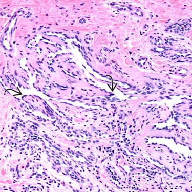
 . The surrounding stroma shows fibrosis and scattered lymphocytes.
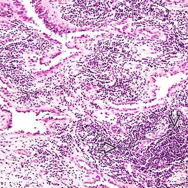
 .
.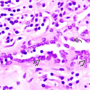
 , and focally enlarged nucleoli
  .
.


