Fig. 4.1
Exanthematous (morbilliform) drug eruption. Erythematous macules and papules, some coalescing
After withdrawal of the offending medication, the rash resolves in 1–2 weeks without sequelae. Treatment is not required, but can provide relief from pruritus. Topical steroids and sedating antihistamines are often used to this end. Systemic corticosteroids are not required in most cases. When the inciting medication is a short-term but essential treatment for the patient (e.g., an antibiotic indicated for a life-threatening infection), it is reasonable to continue the medication while treating the symptoms of the exanthematous eruption. Monitoring for development of systemic symptoms and/or severe adverse reaction while treating through the eruption is advisable, but it does not appear that this type of reaction can progress to a SCAR.
Urticaria and Angioedema
Lesions of urticaria consist of transient, erythetmatous or pale, edematous plaques, sometimes with an annular appearance (Fig. 4.2a). Angioedema may accompany urticarial drug eruptions and represents rapidly progressive edema of the dermis and subcutaneous tissues (Fig. 4.2b). Treatment of drug-induced acute urticaria without angioedema may only require the application of cooling and antipruritic lotions (menthol- or phenol-containing) after removing the suspected medication. For patients requiring systemic medications, it is reasonable to start with a non-sedating antihistamine given daily until the rash clears. If this is not sufficient, then a classic (sedating) antihistamine can be added or substituted. Corticosteroids are generally not indicated.

Fig. 4.2
(a) Wheal of acute urticarial. Erythematous, edematous, annular appearing plaque. (b) Angioedema of the lower lip
Angioedema accompanies urticarial in 50 % of cases and may be associated with anaphylaxis. In the setting of anaphylaxis or angioedema that compromises the airway, subcutaneous epinephrine is indicated in addition to antihistamines. Systemic corticosteroids may also be administered. Strict avoidance of the offending medication in the future is essential, making patient education a major goal in the management of angioedema with or without anaphylaxis.
Fixed Drug Eruption
Fixed drug eruptions appear as round to oval sharply demarcated erythematous and edematous plaques, sometimes with a dusky center that may become bullous (Fig. 4.3). The lesions appear 1–2 weeks after an initial exposure and within 24 h upon re-exposure. Lesions typically recur at the same sites with subsequent eruptions, but new sites may appear with recurrences. Generalized fixed drug eruptions can occur rarely and may mimic Stevens Johnson Syndrome. Lesions favor the lips, hands, feet and genitalia, but may occur anywhere. Treatment is not required, but if lesions are symptomatic, then antihistamines and topical corticosteroids can be used. For patients with generalized bullous FDE, a short course of systemic steroids can be used.
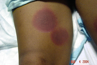
Fig. 4.3
Fixed drug eruption. Round, well-demarcated, erythematous plaques, one with a dusky center
Phototosensitive Drug Eruptions
There are two types of photosensitive drug eruptions: phototoxic and photoallergic. Both are initiated by the combination of the causative medication and ultraviolet A light (UVA) exposure. Phototoxic eruptions present as an exaggerated sunburn, possibly with blisters. Prevention is the best treatment since these reactions are often predictable based on the medication prescribed. Photosensitive eruptions should be treated like sunburns, with cool compresses, emollients, and analgesics. If blisters are present, then wound care to prevent infection should be emphasized.
Photoallergic drug reactions are eczematous, pruritic eruptions in sun-exposed areas. This reaction can be treated with topical corticosteroids. For severe cases, a tapering 2- to 3- week course of prednisone may be required. For both types of reactions, discontinuation of the medication should be considered during the eruption. If the medication must be restarted or continued, then the patient should be educated on photoprotection, including the use of broad-spectrum sunscreens.
Drug-Induced Hypersensitivity Vasculitis
Drug-induced hypersensitivity vasculitis presents with palpable purpura and possibly a maculopapular rash with a temporal relationship to an offending drug (Fig. 4.4). Noncutaneous organ involvement is possible and should be assessed for in the examination and workup. Other findings may include urticaria, arthralgia, fever, lymphadenopathy, low serum complement levels, and elevated erythrocyte sedimentation rate or C-reactive protein. Biopsy of the skin reveals leukocytoclastic vasculitis. Immunofluorescence microscopy can reveal IgM, IgG, or IgA in cases that are induced by a drug, and therefore we do not routinely recommend performance of such testing when there is an obvious drug that was a presumed cause. Infections can cause a similar clinical scenario and should be excluded as a cause where possible. If the leukocytoclastic vasculitis is due to a medication, then discontinuation of the suspect medication should lead to resolution of signs and symptoms within a few weeks. If the vasculitis is severe or does not improve after stopping the inciting medication, the patient may be treated with non-steroidal anti-inflammatory drugs, colchicine, dapsone, or systemic steroids. For cases with progressive or severe systemic disease, immunosuppressive, steroid-sparing agents can be considered.
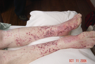
Fig. 4.4
Leukocytoclastic vasculitis. Petechiae and palpable purpura
Acute Generalized Exanthematous Pustulosis
Acute generalized exanthematous pustulosis (AGEP) is caused by drugs in the majority of cases, and occurs hours to days after the offending medication is started. It presents as tiny, numerous, non-follicular-based pustules occurring on a background of erythema (Fig. 4.5). The reaction typically begins in the flexural areas but can become diffuse. Fever may accompany an AGEP reaction, along with leukocytosis and neutrophilia. Prognosis is excellent and the course is typically self-limiting, resolving within 1–2 weeks after stopping the medication. Management beyond withdrawal of the causative medication is supportive care. Pustules typically resolve with superficial desquamation forming a collarette of scale. Coalescence of pustules, however, can result in a clinical picture similar to toxic epidermal necrolysis. In cases with extensive desquamation, wound care should be emphasized to avoid infection, and patients should be monitored for electrolyte imbalances. This may include non-adhesive and/or antiseptic dressings. In mild cases, symptoms and pruritus can be controlled with topical steroids, followed by emollients during the post-pustular phase. Drugs that induce AGEP can produce a positive patch test result with pustules at the site of application. A positive patch test can be confirmatory, but a negative patch test does not rule out a medication as a potential cause.
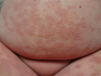
Fig. 4.5
Acute generalized exanthematous pustulosis (AGEP). Non-follicular-based minute pustules on an erythematous base
Drug Reaction with Eosinophilia and Systemic Symptoms
Drug reaction with eosinophilia and systemic symptoms (DRESS) is a potentially life-threatening eruption with a long (2- to 8-week) latency following drug exposure. Skin findings begin with a morbilliform eruption that progresses to involve over 50 % body surface area with confluent, infiltrative plaques (Fig. 4.6). Exfoliative dermatitis, scale, purpura, bullae secondary to edema, and pustules may also be present. Additional signs and symptoms may include facial edema, fever, lymphadenopathy, and malaise. After excluding other causes of the clinical presentation (infection, lymphoma, autoimmune disease), workup for internal organ involvement should include a complete blood count, comprehensive metabolic panel (including liver function tests), urinalysis, pulse oximetry, creatine kinase, troponins, and electrocardiogram. Further testing may be warranted based on signs, symptoms, and results of these screening tests, as shown in Table 4.1.
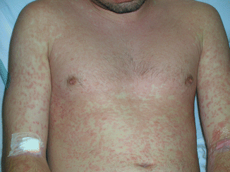
Fig. 4.6
Morbilliform eruption in a patient with DRESS syndrome. Erythematous macules and papules coalescing into plaques
Table 4.1
Recommended investigations for patients suspected of having DRESS
Recommended tests for suspected DRESS | Tests to exclude alternative diagnoses | Further investigations (based on patient presentation) |
|---|---|---|
CBC | Blood cultures | CT scan |
CMP (esp creatinine) | Hepatitis profile | Abdominal ultrasound |
Liver function tests | Antinuclear antibodies | Endoscopy |
Urinalysis | Lymph node biopsy if concern for malignancy | Biopsy of affected organs/sites |
Creatine kinase, troponin | – | PT/INR |
Pulse oximetry | – | – |
EKG | – | – |
Inflammation markers (CRP, ESR) | – | – |
PCR for HHV 6, HHV 7, CMV, EBV | – | – |
After identifying and discontinuing the suspect medication, the treatment for DRESS depends on the severity and organ systems involved. For patients with skin disease and minor liver transaminase elevations, treatment can be supportive and aimed at alleviation of symptoms. Topical steroids and emollients with or without wet wraps can be used for skin discomfort and pruritus. If the patient has extensive exfoliative dermatitis, fluid and electrolyte monitoring and correction may be needed. Systemic corticosteroids should be reserved for patients with pulmonary or kidney involvement (liver disease has not been shown to respond to systemic steroids). Doses of 0.5–2 mg/kg/day of prednisone (or equivalent dose of another systemic glucocorticoid) are recommended until disease stabilizes or improves. Systemic steroids should be tapered slowly over an 8- to 12-week period to avoid relapse. Monitoring for resolution of systemic involvement should be performed at regular intervals during the taper, and shortly afterwards, since relapse is possible. Treatment with steroid-sparing immunosuppressive agents and intravenous immunoglobulin (IVIG) has been reported, however, their use is not routinely recommended. Patients with progressive, severe liver involvement may require transplantation.
Stay updated, free articles. Join our Telegram channel

Full access? Get Clinical Tree


