Fig. 38.1
Angioedema in a young boy. Drugs should always be considered as an underlying etiology
Histopathology
Acute urticaria consists of interstitial edema, dilated venules with endothelial swelling, and minimal inflammatory cells. Chronic urticaria consists of interstitial edema of the dermis and a perivascular and interstitial inflammatory infiltrate with lymphocytes, neutrophils, and eosinophils.
Management
Antihistamines and cessation of the suspected agent(s) is necessary. Oral steroids and antihistamines can be administered for symptomatic relief as needed.
Serum Sickness-Like Reaction (SSLR)
SSLR is a type III hypersensitivity reaction that is more common in infants and children than adults. It is due to the deposition of antigen-antibody complexes in the tissue with subsequent activation of the complement pathway. In contrast to true serum sickness reactions, these lack immune complex deposition, vasculitis, renal lesions, and hypocomplementemia. SSLR presents as an urticarial, ecchymotic, or morbilliform eruption accompanied by fever, lymphadenopathy, arthralgia, eosinophilia, proteinuria, and splenomegaly. This reaction typically occurs within 3 weeks of drug initiation.
SSLR is associated with a variety of medications, including but not limited to penicillins, tetracyclines, sulfonamides, cefprozil, macrolides, itraconazole, griseofulvin, and biological agents (listed below). Studies have suggested that the risk is greater with cefaclor than other antibiotics. Although SSLRs are self-limited, the lesions persist more than 24–36 h and resolve 1–6 weeks after drug cessation.
Drug-Induced Serum Sickness-Like Reaction
Buproprion
Cefazolin
Cefprozil
Cefuroxime
Ciprofloxacin
Clopidogrel
Efalizumab
Fluoxetine
Griseofulvin
Immunoglobulin
Infliximab
Insulin
Itraconazole
Macrolides
Meropenem
Mesalamine
Minocycline
Omalizumab
Penicillin
Rifampicin
Rituximab
Streptokinase
Sulfonamide
Tetracycline
Histopathology
A superficial-to-mid dermal edema with superficial perivascular lymphocytic infiltrate is present without evidence of vasculitis.
Management
Treatment is symptomatic, with removal of the offending drug, antihistamines, and NSAIDs (for arthralgias). Oral prednisone can also be used. Cross-reactivity of cefaclor or cefprozil with other cephalosporins or beta-lactams is rare, and other cephalosporins do not need to be avoided. This is controversial, however, and some clinicians will strongly dissuade the use of any beta-lactam after a cephalosporin-induced SSLR.
Exanthematous Reactions
Maculopapular Exanthems
Also know as morbilliform, simple exanthematous, or scarlatiniform exanthems, these drug reactions are very common, arguably the most common reaction in children, occurring in 1–5 % of first-time drug users. Thought to be a type IV T-cell mediated hypersensitivity reaction, these eruptions frequently occur with penicillin intake and concomitant Epstein-Barr virus (EBV) infection. Patients with HIV and/or bone marrow transplantation are at increased risk. Associated drugs include penicillins, cephalosporins, sulfonamides, and anti-seizure medications, as listed here:
Drug-Induced Maculopapular Reaction
Antiepileptics
Cephalosporin
Penicillins
Sulfonamides
Cutaneous manifestations may occur 6–12 h after drug intake, but typically begin within 1–2 weeks. The maculopapular exanthem is pruritic and morbilliform in nature, with symmetric macules and papules, starting on the trunk and spreading to the face and extremities. No pustules or blisters are present. Erythroderma, palmoplantar involvement, and fever may occur. Mucous membranes are typically spared. Before resolving, the rash becomes hyperpigmented and red-brown, and then ultimately desquamates after approximately 2 weeks.
Histopathology
The findings are relatively non-specific, with a superficial perivascular lymphocytic.
Differential Diagnosis
Viral exanthems can be practically identical in presentation and histopathologically and viral titers can be helpful. Clinical features can aid in the exclusion of acute graft-versus-host disease, toxic shock syndrome, scarlet fever, Kawasaki disease, and juvenile arthritis.
Management
Symptomatic treatment is indicated, as the reaction is typically self-limited. The risks and benefits of continuing or discontinuing the medication(s) should be weighed carefully. If the drug cannot be stopped, close monitoring is best. Reactions can dissipate even when the causative drug is continued. For the pruritus, antihistamines, emollients, and topical corticosteroids can help. These reactions do not progress to more severe, life-threatening reactions, but it is best to assess the likelihood of an early evolving severe reaction. Worrisome signs include bullae, facial edema, fever, mucosal involvement, and positive Nikolsky sign. With re-challenge of the offending medication(s), a reaction may appear quickly, often within 72 h.
Drug Rash with Eosinophilia and Systemic Symptoms (DRESS)
Also known as Drug-Induced Hypersensitivity Syndrome (DIHS) or drug-induced delayed multiorgan hypersensitivity (DIDMOHS; see separate chapter on Drug-induced Delayed Multiorgan Hypersensitivity), DRESS syndrome is a drug reaction that begins several weeks (median time 22 days) after drug exposure and can be life-threatening. Diagnosis is based on the presence of fever, rash, systemic symptoms, and blood eosinophilia. DRESS typically occurs on the first drug exposure, within 1–6 weeks, and has an incidence of 1:3000 exposures. DRESS can lead to mortality in 10 % of affected patients. There is a genetic predisposition in individuals with certain HLA types, and first-degree relatives have a higher risk of developing similar drug reactions.
According to the European Registry of Severe Cutaneous Adverse Reaction study group, antiepileptics were involved 35 % of the time, allopurinol 18 %, sulfonamides 12 %, dapsone 12 %, and other miscellaneous antibiotics 11 % of the time (see the list below). Of the antiepileptics, the most commonly implicated drugs are the aromatic compounds, including carbamazepine, phenytoin, and phenobarbital. Minocycline also has a high reported incidence.
Drug-Induced DRESS Syndrome
Abacavir
Allopurinol
Atenolol
Azathioprine
Captoril
Carbamazepine
Clomipramine
Dapsone
Diltiazem
Gold salts
Isoniazid
Lamotrigine
Mexiletine
Minocycline
NSAIDs
Oxicam
Phenobarbitone
Phenytoin
Sulfonamides
Trimethoprim
High fever is the first manifestation of disease (>38 °C), quickly followed by a violaceous rash, cervical lymphadenopathy and pharyngitis. The rash is present in 95 % of patients with DRESS and may last several weeks. It begins on the face, often peri-orbitally, and upper trunk, and subsequently spreads caudally. The rash can have varied presentations including a morbilliform exanthem (80 % of cases), erythroderma (10 % of cases), exfoliative dermatitis, vesicobullous eruption, pustular eruption, or targetoid lesions. Mucosal sites are involved in approximately 25 % of cases, including the mouth, lips, throat, and genitalia. Facial edema is present in 25 % of cases. Systemic involvement includes lymphadenopathy, hepatomegaly (50 % of cases), myocarditis, lung disease, gastrointestinal symptoms, and endocrine abnormalities. Thyroiditis is a delayed manifestation, often presenting several months after disease onset.
DRESS is thought to be due to a delayed T-cell mediated hypersensitivity reaction, but the exact cause remains unknown. Re-challenge with the offending medication causes return of fever and erythroderma within hours. Of note, the antiepileptics can have significant cross-reactivity; therefore, if there is a reaction to carbamazepine, phenobarbital, or phenytoin, the patient should avoid all three.
Laboratory Evaluation
A complete blood count often shows an atypical lymphocytosis and eosinophilia. Other recommended tests include coagulation panel, viral serologies (hepatitis B, C, EBV, HHV-6), liver and renal function tests, muscle enzymes, thyroid function tests, and/or glucose levels.
Histopathology
A sparse perivascular inflammatory infiltrate with lymphocytes and eosinophils typifies the morbilliform rash. Eosinophils may be absent. Vacuolization of the basal layer and rare apoptotic keratinocytes may be seen. These features are all relatively non-specific, as the histology of DRESS syndrome is not pathognomonic.
Differential Diagnosis
Viral exanthem, erythema multiforme, fixed drug eruption, TEN.
Management
Timely withdrawal of the medication(s) is crucial. In patients with systemic involvement, prednisone (1–2 mg/kg/day) is usually indicated. For symptomatic relief, antihistamines and topical corticosteroids are beneficial.
Pustular Reactions
Acneiform Reactions
Drug-induced acneiform reactions are characterized by monomorphic follicular pustules and papules that affect both acne- and non-acne-prone areas (extremities) and heal sans scarring (Fig. 38.2). Mucosal changes are not present. Medications that have been shown to cause this type of reaction include corticosteroids, lithium, androgens, iodides, bromides, adrenocorticotropic hormone, androgens, actinomycin D, phenytoin, and isoniazid. Newer medications such as epidermal growth factor receptor inhibitors (EGFR/HER1) cetuximab, erlotinib, and panitumumab, also have a high frequency of acneiform eruptions, as listed below. The dose and length of therapy with corticosteroids is directly proportional to the risk of developing an acneiform reaction and those with a history of severe acne are at increased risk.
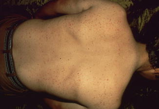
Fig. 38.2
Inflammatory acneiform papules over the back of a teenage boy who had been on long-term systemic corticosteroids
Drug-Induced Acneiform Eruptions
Actinomycin D
Adrenocorticotropic hormone
Androgens
Bromides
Corticosteroids
EGFR receptor inhibitors (cetuximab, erlotinib, panitumumab)
Iodides
Isoniazid
Lithium
Phenytoin
Management
Benzoyl peroxide, in addition to topical or oral antibiotics, as well as topical tretinoin cream, can be helpful as well as discontinuation of the offending medication(s).
Acute Generalized Exanthematous Pustulosis
AGEP is a serious cutaneous reaction with a reported incidence of one to five cases per million persons per year (also see the chapter on AGEP). AGEP rarely affects children, but when so, it is associated with viral (Coxsackie, parvovirus B19, cytomegalovirus, enterovirus) and bacterial (Mycoplasma pneumonia, Chlamydia pneumoniae) infections, as well as vaccinations.
The most commonly implicated drugs include penicillin, cephalosporins (cefixime), vancomycin, clindamycin, acetominophen, paracetamol, bufexamac, cytarabine, and labetalol. Mercury exposure has also been a reported cause of AGEP, as shown here:
Drug-Induced AGEP
Acetominophen
Alphonamides
Amoxicillin
Ampicillin
Bufexamac
Cefixime
Clindamycin
Cytarabine
Diltiazem
Hydroxychloroquine
Labetalol
Mercury
Paracetamol
Penicillin
Quinolones
Terbinafine
Vancomycin
Clinically, AGEP presents with diffuse mildly pruritic and edematous erythema of the intertriginous areas. Subsequently, numerous sterile non-follicular pustules develop. This reaction occurs within hours of drug intake. Fever is usually present but afebrile cases have been reported. Mucous membranes are involved in 20 % of cases and extracutaneous involvement is rare.
Studies have demonstrated AGEP is a drug-specific process mediated by CD4 T-cells, which release GM-CSF and IL-8/CXCL8 cytokines, the latter of which is a potent neutrophil chemoattractant.
Laboratory Evaluation
Peripheral leukocytosis is common, with a neutrophil count over 7000/μL.
Histopathology
Subcorneal or intraepidermal pustules, superficial papillary edema, and a lymphohistiocytic perivascular inflammatory infiltrate are present. Scattered eosinophils and neutrophils can be seen. Single-cell keratinocyte necrosis or vasculitis may be seen.
Differential Diagnosis
DRESS syndrome, pustular psoriasis, leukocytoclastic vasculitis, and subcorneal pustular dermatosis. Differentiation histopathologically may be impossible.
Management
After the offending agent(s) are removed, the reaction should resolve within a couple of weeks. Fine desquamation without scarring may occur. Anti-histamines and a short course of oral corticosteroids (1–2 mg/kg/day) can be used for symptomatic relief of pruritus.
Vesicobullous Eruptions
Fixed Drug Eruption
Fixed drug eruption (FDE) is a drug reaction that classically occurs in the same location with every re-administration of a particular drug (also see the Fixed Drug Eruption chapter). FDEs are relatively common in children, accounting for 10–14 % of ADRs. It may be very difficult to determine the causative drug. The most common drugs implicated in fixed drug eruptions are listed here:
Fixed Drug Eruptions
Acetylsalicylic acid
Amoxicillin
Barbiturates
Co-trimoxazole
Methylphenidate
NSAIDs
Paracetamol
Phenylbutazone
Phenytoin
Pseudoephedrine
Sulfamethoxasole
Teicoplanin
Tetracycline
Trimethoprim
Vancomycin
Clinically, there is a mucocutaneous distribution of pruritic or painful, well-circumscribed and edematous, round red-to-purple patches. They can be solitary or multiple. Vesicles and blisters are variably present. Lesions heal with pigmentary alteration, often darkly hyperpigmented. The most common sites of involvement include lips, trunk, legs, arms, and genitals. Lesions occur within 14 days of original medication assault, and the latency period decreases with subsequent administrations.
The pathogenesis is unclear, but intraepidermal cytotoxic CD8+ T cells most likely release pro-inflammatory cytokines with drug administration. Expression of intercellular adhesion molecules (ICAM) is seen specifically in lesional epidermis, which may explain the sharp localization and circumscription of the lesions. With drug re-challenge, a flare is usually noticed within 1–8 h.
Histopathology
Hydropic degeneration of basal layer keratinocytes, lymphocytic lichenoid infiltrate, and superficial dermal melanophages are present. Scattered necrotic keratinocytes are also seen. Bullae can be seen, as well as extensive confluent epidermal necrosis. Detachment of the epidermis does not have to occur for necrosis to be present. Histologic distinction from erythema multiforme and TEN is not always achievable.
Management
Mostly supportive, but topical corticosteroids may be helpful.
Pseudoporphyria
Pseudoporphyria is a photosensitive bullous skin disease clinically and histopathologically indistinguishable from porphyria cutanea tarda (PCT), but lacks a biochemical porphyrin abnormality. Excessive sunlight, UVA exposure, and certain drugs are supposed to be etiological factors of pseudoporphyria. These drugs include ciprofloxacin, furosemide, tetracycline, dapsone, pyridoxine, NSAIDs (especially naproxen), and oral contraceptives.
Clinically, pseudoporphyria presents with skin erythema, fragility, blistering, and scarring on photo-exposed areas, with a predilection for the face, dorsal hands, and extensor surfaces of the legs. Milia, waxy skin, and hypertrichosis, which are seen in erythropoetic porphyria (EPP) and PCT, are absent in drug-induced porphyria. Lang et al. reported that 12 % of children taking naproxen for juvenile arthritis developed pseudoporphyria. Unlike PCT, no abnormality in porphyrin metabolism has been identified in these cases.
Histopathology
Cell-poor blisters with festooning are present and resemble PCT histologically.
Management
In drug-induced pseudoporphyria, discontinuation of the suspected drug is recommended and necessary. It can take months after discontinuation of the offending drug for resolution of blister formation. Sun protection is advised for all patients.
Drug-Induced Linear IgA Bullous Dermatosis (LABD)
Although rare in children, reports suggest that almost two-thirds of LABD cases may be drug induced. In these cases, LABD presents as an idiopathic autoimmune subepidermal blistering disease. Implicated drugs include antibiotics (frequently vancomycin), NSAIDs, and diuretics. Other drugs include penicillin, cephalosporins, ACE inhibitors, phenytoin, sulfonamides, and rarely amiodarone, atorvastatin, carbamazepine, cyclosporine, furoseminde, gemcitabine, glyburide, GCSF, influenza vaccination, lithium, rifampin, PUVA, somatostatin, verapamil and vigabatrin. In children, most cases are secondary to infections and/or drugs. After the drug is discontinued, the prognosis is excellent. Of note, there have been few reports of increased morbidity secondary to pruritus.
Stay updated, free articles. Join our Telegram channel

Full access? Get Clinical Tree


