SCR presents as single papular or nodular cutaneous lesion


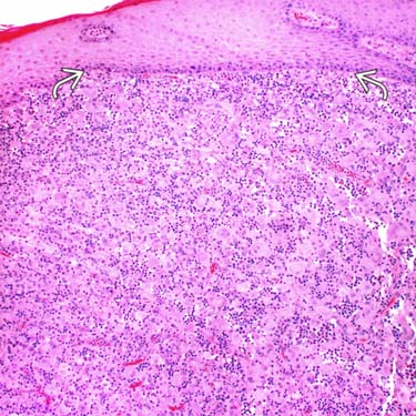
Low magnification of multicentric reticulohistiocytosis (MCR) shows a nodular proliferation of enlarged histiocytic cells in the dermis. There is a very thin grenz zone
 , and the epidermis shows flattening of the rete ridges.
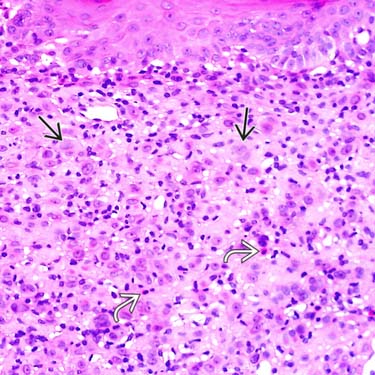
Higher magnification of MCR shows a proliferation of numerous, enlarged, histiocytic-appearing cells
 associated with a background mixed inflammatory infiltrate containing scattered eosinophils
  .
.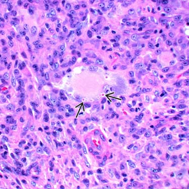
High magnification of a cellular MCR shows a very large, central multinucleated cell containing fragments of phagocytosed erythrocytes
 .
.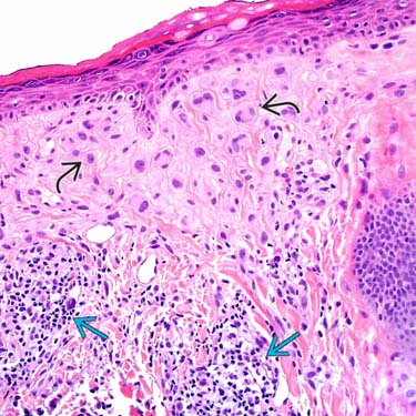
A hypocellular example of MCR shows a superficial, dermal-based proliferation of large epithelioid histiocytes
 , many of which are multinucleated, and a mild associated lymphocytic infiltrate
  .
.CLINICAL ISSUES
Epidemiology

Presentation
• Multiple nodules & arthropathy
 Most cases present with multiple cutaneous/mucocutaneous papulonodules & severe arthropathy, other visceral symptoms


Most cases present with multiple cutaneous/mucocutaneous papulonodules & severe arthropathy, other visceral symptomsStay updated, free articles. Join our Telegram channel
Full access? Get Clinical Tree
