2
Introduction to the Head and Neck
The head and neck are two distinct anatomical regions of the body, but they have a related nerve and blood supply.
Head
The head is a highly modified structure with several important functions. It houses and protects the special sense organs—the eyes, ears, nose, tongue, and related structures. The skull is specially adapted to enclose, support, and protect the brain (Fig. 2.1). It has numerous foramina for cranial nerves and vascular structures to pass into and out of the cranium, contains cavities that carry out some of the functions of the upper gastrointestinal and respiratory tracts (e.g., oral and nasal cavities), and provides a foundation for the face. Anatomically, the skull is divided into two main parts:
- The neurocranium houses the brain, forms the base of the skull and cranial vault, and is composed of eight bones—the occipital, sphenoid, frontal, and ethmoid bones; a pair of parietal bones; and a pair of temporal bones.
- The viscerocranium (facial skeleton) contributes to the structure of the orbits and the nasal and oral cavities and provides a foundation for the face; it comprises the mandible and vomer and a pair each of the maxillary, palatine, nasal, zygomatic, lacrimal, and inferior nasal concha bones.
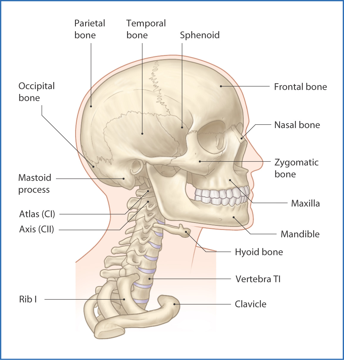
FIGURE 2.1 Bones of the head and neck.
The paranasal sinuses are cavities within the maxillary, ethmoid, frontal, and sphenoid bones that communicate with the nasal cavity through small ostia (openings).
Neck
The head is mobile because the skull is balanced on the flexible bony spine. The neck extends from the base of the skull (a circular line joining the superior nuchal line, mastoid process, and lower border of the mandible) to the chest (sternum, clavicles, spine of the scapula, and spinous process of cervical vertebra CVII). It is a flexible conduit for blood vessels, the spinal cord, and cranial and spinal nerves passing between the head, thorax, and upper limb.
The neck is supported by muscles, ligaments, and the cervical vertebrae, which provide a strong, flexible skeletal framework without sacrificing stability. The seven cervical vertebrae have vertebral foramina (for the vertebral arteries to pass through) within their transverse processes (see Chapter 26). The cervical segment of the vertebral column is strongly supported by numerous ligaments and muscles (both extrinsic and intrinsic). Intermediate parts of the respiratory tract (larynx and trachea), digestive tract (pharynx and esophagus), and endocrine glands (thyroid and parathyroid glands) are located within the neck.
For descriptive purposes the neck is subdivided into anterior and posterior triangles. These two large triangles are further subdivided into minor triangles: submandibular, submental, carotid, muscular, occipital, and omoclavicular (subclavian) triangles (see Chapter 12).
The fascia of the neck is multilayered and encloses the muscles, glands, and neurovascular structures. The relationships between the different fascial layers determine how infection and cancer spread in the neck. The deep cervical fascia subdivides the neck into vascular, vertebral, and visceral compartments. This arrangement allows movement between adjacent structures and compartments and facilitates the surgical approach to specific areas. The investing layer of cervical fascia encircles all structures of the neck by investing the sternocleidomastoid and trapezius muscles, the fascial roofs of the anterior and posterior cervical triangles, and the parotid and submandibular salivary glands. Deep to the investing fascia and surrounding the visceral compartment is the pretracheal layer of cervical fascia; this layer invests the trachea, thyroid and parathyroid glands, and the buccopharyngeal fascia, which extends from the base of the skull and envelops the buccinator muscle and pharyngeal constrictors.
The cervical part of the vertebral column and its contents form the vertebral compartment of the neck and are surrounded by the prevertebral layer of fascia. The brachial plexus passes between the anterior and middle scalene muscles and is enclosed in a prolongation of the prevertebral fascia—the axillary sheath. The suprapleural membrane, which covers the apex of the lungs, is continuous with the prevertebral fascia and continues into the thorax as the endothoracic fascia.
Two special fascial units—the carotid sheaths—extend from the base of the skull to the superior mediastinum. These sheaths enclose the common and internal carotid arteries, the internal jugular vein, and the vagus nerve [X] and are surrounded by the deep cervical lymph nodes (see p. 11).
Muscles
The major muscles of the head and neck are derived embryologically from two major sources:
Stay updated, free articles. Join our Telegram channel

Full access? Get Clinical Tree


