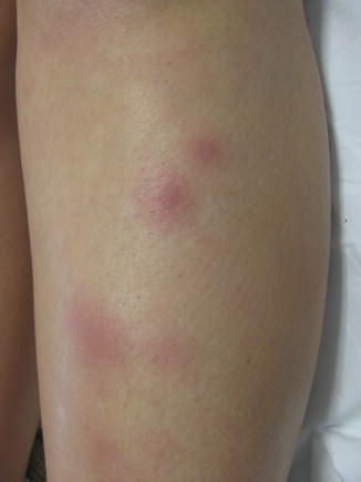
Fig. 11.1
Symmetrically distributed on the anterior shins there are erythematous, warm, indurated plaques that were confirmed to be EN on histopathology

Fig. 11.2
A close-up of the lesions of EN on the left shin
EN as a Drug Eruption
Many medications are associated with EN; common agents include estrogen-containing oral contraceptive pills (OCPs), sulfonamides, penicillin, bromides, iodides, tumor necrosis factor (TNF) inhibitors, and granulocyte colony stimulating factors (G-CSF). There are other, less common, causes of drug-induced EN, as listed here:
Antibiotics (sulfonamides, penicillin)
Bromides
Iodides
TNF inhibitors
Granulocyte colony stimulating factors
Oral contraceptive pills (estrogen containing)
Progesterone (intramuscular)*
Azathioprine*
Vemurafenib*
Propylthiouracil*
Interferon*
*denotes less common cause
Azathioprine (AZA) hypersensitivity reaction can present with EN. A patient had several red-purple nodules on both lower legs 1 week after starting AZA 50 mg daily for bullous pemphigoid (BP), despite having normal thiopurine methyltransferase (TPMT) enzyme activity. Punch biopsy confirmed the diagnosis of EN and there was complete resolution of the lesions within 2 weeks of discontinuing AZA. This is considered an idiosyncratic hypersensitivity reaction to AZA and does not occur commonly.
EN can also be an adverse effect of anti-TNF agents, specifically certolizumab. A patient was being treated for CD with certolizumab (400 mg every 4 weeks); 1 day after her second injection, she noticed a tender, red nodule on her left lateral ankle. Imaging ruled out thrombophlebitis and deep-vein thrombosis (DVT). With each subsequent injection, the patient noticed expanding erythematous plaques on the anterior lower legs that were confirmed histologically to be EN. Once the certolizumab was stopped, the skin lesions started to resolve while the CD flared, supporting the theory that the EN was from the certolizumab rather than the underlying CD.
Vemurafenib is an oral agent used to treat metastatic melanoma; it is an inhibitor of mutated BRAF gene. Similar to other targeted therapies, vemurafenib has been associated with cutaneous adverse events. Two patients who were receiving 960 mg of vemurafenib both developed painful lesions on their arms and legs within 40 days after starting the medication. Biopsy was consistent with EN. A thorough infectious and autoimmune workup was done and was unrevealing, thereby attributing the EN to vemurafenib.
Propylthiouracil (PTU) is a drug used to treat hyperthyroidism. It has various adverse effects, many of which are cutaneous. These cutaneous manifestations include urticaria, pruritus, hair loss, and erythema multiforme. Some of the rare cutaneous findings associated with PTU include antineutrophil cytoplasmic antibody (ANCA)-positive vasculitis, which has a predilection for the distal extremities and the face but can also involve the ears, trunk, and breasts. Pathology of these lesions would show a small vessel leukocytoclastic vasculitis (LCV). There have been few reports of an ANCA-positive erythema nodosum. The incidence of PTU-induced ANCA positivity is 4–6 %. The time between starting PTU and ANCA positivity ranges from 1 week to 13 years. Approximately 20 % patients with positive ANCA develop vasculitis. A patient with hyperthyroidism had been started on PTU 100 mg daily and after 11 months developed warm, tender, red nodules ranging in size from 1 to 5 cm on bilateral, anterior lower extremities. She also had smaller, similar lesions on her fingers, palms, and wrists. Laboratory analysis revealed a normal complete blood count and a positive perinuclear (p)-ANCA at 1:160 dilution, while her proteinase 3 (PR3)-ANCA level was 0. Pathology confirmed the diagnosis of EN and there was no evidence of vasculitis. PTU was stopped and the EN was treated with thalidomide 75 mg daily. Over the next 3 weeks the lesions resolved, and 3 months later the p-ANCA titer decreased to 1:80. At 5 months, thalidomide was stopped. One year later the patient’s skin was clear, with no recurrence of EN.
Stay updated, free articles. Join our Telegram channel

Full access? Get Clinical Tree


