Fig. 7.1
Atypical target lesions with dusky center (typical of people of color) and surrounding erythema in a patient who flares with episodes of genital HSV
Historical Perspective
In 1866, Ferdinand von Hebra described an acute, self-limited, mild skin disease characterized by symmetrically distributed, evolving skin lesions. The lesions presented with an acral distribution and had a tendency for recurrences. For much of the nineteenth century, morphologists had identified many different types of “erythema” and used terms such as “erythema papulatum”, “erythema tuberculatum”, “erythema annulare”, “erythema iris”, “erythema gyratum”, and “herpes iris.” von Hebra concluded that each of these terms represented different stages of the same disease, which he called “erythema exudativum multiforme.” The term multiforme exemplifies not the multiple presentations of EM, but instead multiple morphologic stages of the same lesions.
Following the original description of the disease, the term erythema multiforme has been used to describe various diseases, many only minimally resembling von Hebra’s original description. In 1922, Stevens and Johnson described a disease in two boys with acute febrile illnesses and skin lesions somewhat resembling those of EM, along with associated stomatitis and severe purulent conjunctivitis (Fig. 7.2). Stevens and Johnson recognized that this was distinct from von Hebra’s disease and coined “a new eruptive fever with stomatitis and ophthalmia,” which by the 1940s became known as Stevens-Johnson syndrome (SJS). In 1950, Thomas et al. recognized a distinction between the two diseases, and used EM minor to characterize von Hebra’s classic mild disease, and EM major to refer to a more severe disease, consistent with SJS. In 1956, Lyell described toxic epidermal necrolysis (TEN), a more severe disease with extensive skin sloughing. EM minor, EM major, SJS, and TEN were considered to be part of the same disease spectrum, with each term describing a different level of severity.


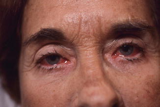
Fig. 7.2
Purulent conjunctivitis in patient with typical target lesions of erythema multiform on areas of the skin not seen in the figure
Until the 1980s and 1990s, the terminology surrounding EM became even more muddled. As mentioned previously, EM major and SJS were often used synonymously. Other literature defined EM as only involving one mucosal surface, whereas SJS involved at least two. Others classified the diseases based on etiology. Such inconsistencies have made gathering meaningful epidemiologic and etiologic data extremely difficult.
Current Classification of Erythema Multiforme
By the 1980s, researchers began to further characterize EM, SJS, and TEN. Howland et al. noted that EM minor, using a definition close to that described by von Hebra, was frequently associated with HSV. This disease tended to be less severe than sulfonamide-associated disease, which had more widespread lesions and increased mucosal involvement. Of note, the authors defined the two presentations as EM minor and EM major.
In 1993, recognizing the confusing terminology, Bastuji-Garin et al. proposed a classification scheme to differentiate EM, SJS, and TEN. Hospitalized patients with suspected erythema multiforme, SJS, or TEN were examined. Four clinical patterns emerged: typical targets, raised atypical targets, flat atypical targets, and macules with or without blisters. Additionally, the percentage of body surface area (BSA) of detached or detachable epidermis was calculated. Five diagnostic categories were proposed:
Bullous EM: detachment below 10 % of the BSA plus localized typical targets or raised atypical targets
SJS: detachment below 10 % of the BSA plus widespread macules or flat atypical targets
Overlap SJS/TEN: detachment between 10 and 30 % of the BSA plus widespread macules or flat atypical targets
TEN with spots with or without blisters: detachment above 30 % of the BSA plus widespread macules or flat atypical targets
TEN without spots: detachment above 10 % of the BSA with large epidermal sheets and without any macule or target
Both dermatologists and non-dermatologists used this classification scheme, and were able to successful classify lesions with 68–100 % concordance. Furthermore, the authors concluded that EM can be clinically distinguished from SJS and TEN based on morphology, distribution, and etiology.
Using similar classification, Assier et al. confirmed the finding that EM and SJS could be distinguished based on clinical pattern. They also provided evidence that etiologic agents for EM and SJS are distinct, with EM being related to herpes and SJS being more related to drugs. Subsequently, a case-control prospective study with 552 patients confirmed the distinction between EM and SJS, and stated that SJS and TEN are the same disease, with TEN being more severe. This study classified EM as having typical targets, raised atypical targets in a localized, acral pattern with less than 10 % blister involvement. Notably, no distinction was made between EM minor and EM major in these studies. Today, the terminology EM minor and EM major are still in use, but now refer to EM without mucosal involvement and with mucosal involvement, respectively.
Epidemiology
Reported prevalence rates of EM are typically <1 %. However, a paucity of research exists on EM prevalence. In addition to difficulties in classification, the acute nature of the condition and a lack of a reporting registry also contribute to scant epidemiologic data. EM typically affects young adults who are 20–40 years old, but children and the elderly can be affected. Over one-third of cases may be recurrent, and recurrence is even more common in HAEM. Mortality rates for EM are not well reported, but are thought to be low. Conversely, SJS and TEN have mortality rates of approximately 5 and 30 % respectively.
Presentation and Characteristics
The classic presentation as described by von Hebra remains the most important clue for diagnosis of EM. The classical form of EM arises 1–14 days following an episode of herpes labialis or herpes genitalis. Following a period of either absent or mild prodrome, symmetrically distributed lesions develop on the extensor surfaces of the extremities, commonly the dorsal aspects of the hands. The lesions evolve to become characteristic targetoid lesions, which last from 1–4 weeks and resolve in a self-limited fashion. The following discussion will focus on the mild disease most consistent with the disease originally described by von Hebra.
Prodromal Symptoms
EM cutaneous findings are rarely preceded by prodromal symptoms, and when present, these symptoms tend to be mild. When prodromal symptoms occur, fever, malaise, headache, cough, rhinitis, sore throat, myalgia, arthralgia and nausea occur 7–14 days before cutaneous lesion development. Whether the prodromal symptoms are a result of the EM disease process itself, or associated with underlying cause (e.g., HSV infection) can be difficult to distinguish.
Morphology
The earliest cutaneous manifestations are round, erythematous macules, which quickly evolve to papules, which may be surrounded by an area of blanching. At this point, the lesions can resemble insect bites or urticarial hives. Lesions then enlarge and develop concentric alterations in morphology in color. Lesions generally range from 2 to 20 mm in size. The central area of the lesion gradually darkens, and either a central blister with a necrotic blister roof, an area of epidermal necrosis without a blister, or an area of crusting develops. This central area may be beefy red, white, yellow, or gray with a darker gray-to-blue rim of color at the edge. Immediately surrounding the area of epidermal necrosis is a dark red, inflammatory zone, which is surrounded by a lighter color, edematous ring. At this point, the lesions are described as targets, or iris lesions, due to their three concentric zones: the central dusky zone, the pink edematous zone, and the peripheral red ring. In individuals with more pigmented skin, the entire area of central necrosis may be dark gray. The lesions may become more complex, and may coalesce, develop central erosions or crusting, or develop central clearing. The inflammatory process in an individual area may remit, and relapse at a later time, further diversifying the appearance. Patients may also have multiple stages of lesions at any one time. The lesions typically heal without scarring, but post-inflammatory hypo- (Fig. 7.3) or hyper-pigmentation may occur, especially in more pigmented skin.


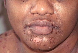
Fig. 7.3
Residual areas of oval hypo-pigmentation around the mouth in a patient with erythema multiforme due to lisinopril. The white areas should resolve, but it may take 4–6 months
Cutaneous Distribution
Lesions typically present symmetrically, with a predilection for the dorsal aspects of the hands and extensor surfaces of the extremities. Hundreds of lesions may be present. Other areas of involvement, although less frequent, are skin of the palms, soles, flexural aspects of the extremities, neck, perineum, ears, and face. The lesions can spread first from extensor surfaces to flexor surfaces, and then centripetally, but involvement of the trunk is less common and less pronounced. The isomorphic phenomenon, also known as koebnerization, and photoaccentuation are thought to play a role in the cutaneous distribution of lesions. Of note, koebnerization is only thought to occur prior to cutaneous eruption, and once skin lesions are present, the phenomenon no longer occurs.
Mucous Membrane Lesions
Although von Hebra’s original description of EM did not involve the mucous membrane, significant literature describes such an association. Estimates on mucous membrane involvement range from 25 to 70 %. When mucosa is involved, it usually occurs simultaneously with, but may occur before or after, cutaneous manifestations. Mucous membrane involvement may rarely occur in the absence of cutaneous involvement. The oral mucosa is most commonly involved, with the labial mucosa, buccal mucosa, non-attached gingivae, and vermillion lip being common locations (Fig. 7.4). The lesions range from diffuse oral erythema and edema to multifocal superficial ulcerations. Other reported mucous membranes include ocular, genital, upper respiratory, and pharyngeal mucosa.


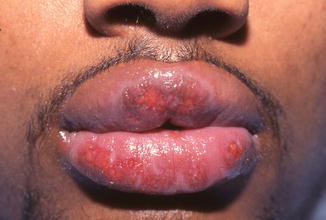
Fig. 7.4
Erosions over the lip of erythema multiforme secondary to ibuprofen
Associated Symptoms
EM tends to be localized to the skin and mucous membranes with few systemic symptoms. When symptoms occur, patients complain of mild malaise, itching and burning over the skin, and pain associated with mucosal erosions. Fever, myalgia, arthralgia, and intense headache are rarely present.
Course of Illness
New lesions usually occur over 3- to 5-day periods, but may also erupt over 1–2 weeks. In this “eruptive” phase, lesions may occur in groups. Lesions tend to heal in less than 4 weeks. Lesions do not scar, but may lead to hypo- or hyper-pigmentation.
Complications
Complications in EM are typically minor. Oral involvement may lead to decreased oral intake, which can lead to dehydration. More serious complications such as keratitis, conjunctival scarring, uveitis, or even permanent vision loss, have been reported. Also reported are esophagitis, esophageal strictures, and upper airway lesions leading to pneumonia. Whether or not these more serious complications are truly a result of EM is debatable. Instead, these cases could have been misclassified as the more severe SJS and TEN.
Atypical Presentations
EM lesions are not always classical, and clinical manifestations of EM vary from patient to patient. Atypical lesions are not “targetoid,” but instead only have two concentric zones and have areas of palpable, round, edematous lesions with poorly defined borders.
Drug-Induced Erythema Multiforme (DIEM)
Much of the early literature regarding DIEM included sulfonamide-induced disease, which was characterized by large bullae, widespread disease, severe mucosal involvement, fever, and prostration. These reactions were often termed EM major, and are likely better characterized as SJS. A challenge exists when lesions are associated with a drug exposure, and present with targetoid lesions in an atypical pattern, but do not have mucosal involvement or skin sloughing, and thus do not fit the description of SJS. These lesions may be best classified as an EM-like reaction, distinct from classical EM. When these reactions occur, the lesions are often flat atypical two-zoned targets. In addition to atypical skin lesions, DIEM is more likely to have a flu-like prodrome, is more likely to involve mucous membranes, and is less likely to be recurrent.
Mycoplasma pneumoniae-Associated Erythema Multiforme
While M. pneumoniae continues to be considered an etiologic agent of EM, the clinical presentation is distinct from that of HAEM (Table 7.1), and some authors believe that M. pneumoniae causes SJS, but not EM. M. pneumoniae-associated disease usually affects children and young adults. M. pneumoniae-associated EM is similar to HAEM in that it presents with targetoid lesions roughly half of the time, and that the lesions tend to erupt on the extremities and spread centripetally. In contrast to HAEM, prodromal symptoms are common, and patients have symptoms of fever, chills, sore throat, cough, runny nose, malaise, and myalgia 2 days to 2 weeks prior to rash development. Skin lesions tend to be maculopapular or vesiculobullous, and oral lesions tend to be more pronounced. Other mucosa surfaces are also affected commonly, with reported involvement of the genitalia, urethra, ocular mucosa, and anus. Overall, the disease tends to be more severe than HAEM and more frequently requires hospitalization.
Table 7.1


Comparison between HAEM and EM-like drug reactions
Stay updated, free articles. Join our Telegram channel

Full access? Get Clinical Tree


