Fig. 6.1
Symmetric urticarial drug eruption due to cephalexin over the back. This was acute and discontinuation of the drug resulted in rapid resolution
Antibiotics: penicillins, cephalosporins, macrolides, aminoglycosides, tetracyclines, sulfonamides, vancomycin
Nonsteroidal anti-inflammatory agents: ibuprofen, naproxen sodium
Salicylates
Angiotensin-converting enzyme inhibitors
Antifungal agents: fluconazole, ketoconazole
Opioids: morphine, codeine, meperidine, fentanyl
Dextromethorphan
Betadine (povidone-iodine)
Muscle relaxants: atracurium, vecuronium, succinylcholine, curare
Thrombolytics: alteplase, urokinase
Protamine sulfate
Antineoplastics
Steroids
Progesterone
Polypeptide hormones: insulin, corticotrophin, vasopressin
Enzymes: trypsin, streptokinase, chymopapain
Hydantoins
Sorbitol complexes
Hydralazine
Quinidine
Dextrans
Mannitol
Vaccines
Vitamins
Radiographic contrast agents
Type I hypersensitivity is the pathomechanism proposed for penicillin-induced acute urticaria. It is an immune-dependent mechanism that requires an initial sensitization period, during which no allergic reaction usually occurs. The sensitization results in synthesis of drug-specific immunoglobulin E (IgE) antibodies, the Fc portion of which is then bound by high-affinity IgE Fc receptors (FcεRIs) present on the surface of mast cells. This leaves the antigen-binding site exposed to the extracellular space, ready to bind its antigen. When a parent drug or one of its metabolites binds to drug-specific IgE antibodies anchored within the plasma membrane of mast cells, crosslinking of two adjacent Fc receptors may occur. This sequence activates mast cells, the effector cells in urticaria. Activation incites the degranulation of mast cell contents, releasing histamine and other vasodilators into the extracellular environment. The resulting vasodilation leads to increased vascular permeability and extravasation of intravascular fluid. This sequence forms clinically evident wheals. β-lactam antibiotics are the most common culprits of drug-induced acute urticaria by this immediate hypersensitivity allergy. This mechanism may also play a role in chronic urticaria, discussed later.
Acute urticaria may also be mediated immunologically by a type III hypersensitivity reaction that involves immune-complex activation of the complement cascade. This process begins when immunoglobulins G or M (IgG, IgM) combine with an excess of drug antigen, forming multiple antibody-antigen immune complexes. These immune complexes activate the classical complement pathway, through which anaphylatoxins are produced that act on mast cells and basophils to trigger the release of vasodilators. This phenomenon is referred to as serum sickness and is associated with systemic symptoms, including fever, arthritis, neuritis, and papular rash. The onset is subacute, beginning between 6 days and 3 weeks after ingestion of a culprit drug. A set of drugs known to cause serum sickness is listed here (adapted from PMID 16461991). The urticarial rash resolves several weeks after drug cessation.
Penicillins and cephalosporins
Aspirin
Captopril
Sulfonamides
Phenytoin
Globulin preparations
Para-aminosalicylic acid
Allopurinol
Arsenic and Mercury derivatives
Barbiturates
Furazolidone
Gold salts
Griseofulvin
Halothane
Hydralazine
Methyldopa
Iodides
Penicillamine
Piperazine
Procainamide
Quinidine
Streptokinase
Thiouracils
Non-immunologic pathomechanisms may also elicit an acute-type urticaria. Some drugs act directly on mast cells to increase degranulation of histamine and vasodilatory mediators, without prior sensitization. Drugs that induce urticaria by this mechanism include opiates, codeine, amphetamine, polymyxin B, atropine, muscle relaxants, hydralazine, pentamidine, quinine, and radiocontrast media.
The etiology of drug-induced acute urticaria may be multifactorial in some cases. For example, benign viral illnesses are known to sensitize mast cells and cause acute urticaria. If treated with NSAIDs, infection-related urticaria may be exacerbated by a mechanism discussed hereafter in the context of chronic urticaria.
Chronic Drug-Induced Urticaria
Chronic urticaria differs from acute urticaria by its time course. It is defined as a relapsing, remitting urticaria with lesions reappearing at least twice per week for longer than 6 weeks, usually in the absence of any identifiable cause (Fig. 6.2). The prevalence of chronic urticaria is between 0.5 and 3 %, and is rare in children. Chronic urticaria is usually an idiopathic preexisting condition which may be exacerbated by drugs through similar mechanisms responsible for acute urticaria. Mainly, the non-immunologic pathomechanism is involved, in which drugs worsen chronic urticaria by directly increasing mast cell mediator release. Drugs which may exacerbate chronic urticaria include NSAIDs, codeine, and morphinic agents. About 30–75 % of patients suffering from chronic urticaria may experience angioedema after taking aspirin or NSAIDs.
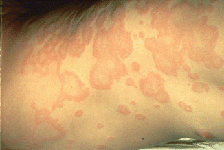
Fig. 6.2
Annular pink plaques with clear centers on the left flank in a patient on multiple medications. The exact etiology was not determined and was chronic in nature
Another pathomechanism implemented in chronic urticaria is a specific pharmacological hypersensitivity related to NSAID intake. This pseudoallergic reaction aggravates chronic urticaria after aspirin or NSAID intake. The prevalence of NSAID-induced urticaria and angioedema in all patients ranges between 0.1 and 0.3 %. Aspirin and NSAIDs are cyclooxygenase (COX) inhibitors that cause pseudoallergic reactions by altering metabolite levels in the arachidonic acid metabolism pathways. Inhibition of COX 1 and COX 2 increases the levels of cysteinyl leukotrienes, leading to vasodilation and edema, causing wheal-and-flare eruptions in patients hypersensitive to these medications. This mechanism is considered a pharmacological side effect rather than a true allergy since no immunogenic phenomenon transpires.
Contact Urticaria
The third type of urticaria related to drug use is contact urticaria. This side effect arises after the application of topical medications. Ordinarily, the more likely adverse reaction caused by topical medications is an eczematous contact dermatitis. However, urticaria can also result as a transient wheal-and-flare eruption. The process may occur within a few minutes to an hour of application, and resolves in fewer than 2 h after removal of the offending topical agent. Contact urticaria develops as a result of both type I hypersensitivity and non-immunologic mechanisms.
The immunologic mechanism may result in generalized urticaria whereas the non-immunologic mechanism usually remains localized to the area of application. Topical antibiotics are common culprits, including penicillin, cephalosporins, gentamycin, neomycin, bacitracin, streptomycin, and chloramphenicol, when administered in topical formulations like ointments, creams, or eye drops. Neomycin causes localized contact dermatitis in addition to contact urticaria. Bacitracin has been reported to cause severe anaphylaxis. Topical anesthetics such as benzocaine and lidocaine within EMLA cream have also been reported to cause contact urticaria. Chlorhexidine, known to cause contact dermatitis and photosensitive reactions, may also cause contact urticaria. Other non-immunologic contact reactions may occur following the handling of substances like ammonium, persulphate (hair perming solution), cinnamic aldehyde, benzoic acid (found in cosmetics and food), and other topical cosmaceuticals (Fig. 6.3). There have been increasing reports of contact urticaria immediate-type hypersensitivities, and severe anaphylactic shock in handlers of these compounds.
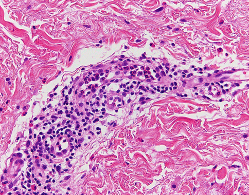
Fig. 6.3
Contact urticaria due to a shampoo. Note that, unlike most cases of contact dermatitis, there is no eczematous or epidermal component, but erythema and edema indicative of an urticarial reaction with dermal edema and vasodilatation
Angioedema
Urticaria is associated with angioedema in 40 % of patients, defined by deep edema within the reticular dermis or subcutaneous tissue. Angioedema should not be viewed as a separate entity, but rather a clinical variant of urticaria at a deeper skin depth (Fig. 6.4). Angioedema differs from urticaria by its ill-defined borders, pale- to skin-color, association with pain and tenderness, and duration of up to 72 h. The phenomenon typically localizes to the lips, eyelids, genitalia, and extremities. Angioedema is a known side effect of angiotensin-converting enzyme (ACE) inhibitors, and occurs between 0.1 and 0.2 % of patients receiving ACE inhibitors. The mechanism involved is due to the potent inhibition of kinase II by the ACE inhibitor. This leads to an increase in bradykinin levels, which results in continued prostaglandin E2 synthesis, leading to vasodilation, increased vascular permeability, and increased fluid leakage into the interstitial space.
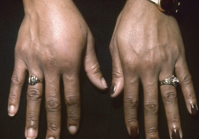
Fig. 6.4
Angioedema of the hand on the left, with an area of large, deep swelling
Presentation
Regardless of the time course and subtype, urticarial wheals present similarly as rapidly emerging, pink to pale plaques with central clearing and surrounding erythematous flare. These transient lesions may be round, oval, or serpiginous in shape, range from a few millimeters to several centimeters in size, and appear anywhere on the body, including the palms, soles, and scalp. They are intensely pruritic and may evoke a burning sensation, but are not usually painful. The distribution is usually random and asymmetric, although chronic urticarial lesions can recur in repeat locations. By definition, lesions appear within minutes to hours, and persist for fewer than 24 h after onset. Upon resolution, there are no residual ecchymoses or discolorations, unless trauma due to scratching was involved. If lesions are painful, or leave residual ecchymoses, then urticarial vasculitis should be considered as a diagnosis.
In acute urticaria, lesions appear within a few hours to days after the drug is taken, and disappear within several days after withdrawal of the drug. If the urticaria is due to an allergy caused by an IgE-dependent immune mechanism, the disease is generalized and potentially fatal, whereas pseudoallergic events are rarely fatal. In chronic urticaria, lesions may resolve from time to time, but repeatedly relapse, preventing the patient’s complete recovery. In contact urticaria, lesions are usually localized to the site of application, but in some cases the urticaria may generalize.
Histopathology
Histological sections of urticaria commonly display dermal edema with mild dilation of dermal blood vessels. Absent are signs of vessel damage and leukocytoclasia. Cellular infiltrations are evident perivascularly with sparse densities of neutrophils, macrophages, eosinophils, and lymphocytes.
Differential Diagnosis
Exanthematous drug eruption: history of drug intake, possible low-grade fever, lesions appearing as fixed maculopapular confluent wheals bilaterally and symmetrically on the trunk
Contact dermatitis (allergic or irritant): carefully-elicited history of offending agent exposure, lesions apparent only at site of contact
Stay updated, free articles. Join our Telegram channel

Full access? Get Clinical Tree


