Diseases commonly associated with or preceding erythroderma a
Dermatologic diseases
Psoriasis
Atopic dermatitis
Pityriasis rubra pilaris
Contact and other types of dermatitis
Dermatophytosis
Ichthyosis
Pemphigus foliaceus
Systemic diseases
Lymphoma: CTCL, Large cell, Hodgkin’s
Leukemia
Carcinoma (of blood vessels, thyroid, lung, esophagus, colon, liver or prostate)
Dermatomyositis
Hepatitis
Renal insufficiency
Immunodeficiency (acquired or congenital)
Histoplasmosis
Toxoplasmosis
Lupus erythematosus
Thyrotoxicosis
Sarcoidosis
Graft-versus-host disease
Hyper-eosinophilic syndrome
Drugs Commonly Associated with Erythroderma
Antihypertensive medications: beta-blockers
Antibiotics: bactrim (trimethoprim-sulfamethoxazole), tobramycin, vancomycin, penicillin, gentamycin, cefoxitin
Antifungals: ketoconazole, griseofulvin
Calcium channel blockers: nifedipine
Proton pump inhibitors: omeprazole
H2 blockers: cimetidine, ranitidine
ACE inhibitors: captopril
Anti-tuberculosis medications
Carbamazepine
Phenobarbital
Paracetamol
Lithium
Plaquenil
Antimalarials
Given this broad-based etiology, evaluation of a patient presenting with the signs and symptoms of erythroderma must include a systematic search for potential etiologic agents, which most often are drugs or pre-existing dermatoses. Other common categories of diseases that may manifest as erythroderma are cutaneous and internal malignancies, such as mycosis fungoides and cancers of the GI tract, respectively. Patients with pre-existing psoriasis can develop erythroderma secondary to the withdrawal of topical or systematic steroid treatments. Thus, it is important to not only identify whether the patient began a new drug regimen recently, but also to assess whether a drug has been ceased recently.
Drug reactions may initially consist of morbilliform, urticarial, or lichenoid rashes that eventually coalesce to present as generalized erythroderma. This pattern may be a tell-tale sign that a drug is the etiologic agent in those cases. Other patterns in the development of erythrodermic symptoms and signs may be telling as well. For example, patients with erythroderma due to underlying malignancy will likely present with a history of gradual onset of skin manifestations, combined with recalcitrance from standard therapeutic approaches and progressive decompensation (as opposed to the rapid decompensation which is more typical of drug-induced erythroderma). Moreover, mucosal involvement suggests the presence of toxic epidermal necrolysis (TEN), which is life-threatening and must be identified and treated immediately. It is usually due to a hypersensitivity-induced reaction to an offending drug. TEN often has cardiac, pulmonary, renal, and ocular complications, which can also help identify the etiology of the disease. Lastly, some herbal preparations or non-traditional medical treatments may cause this disease; however, the body of evidence to support this claim is unclear at this time.
Pathogenesis
The molecular pathogenesis of erythroderma remains elusive. However, a number of key changes have been noted. Table 23.2 lists some of the biochemical changes that have been documented in histologic samples from patients with erythroderma. Note that all of these histologic changes are relatively non-specific and are unlikely, at this time, to be useful for making a diagnosis of erythroderma. However, they may be useful in diagnosing an underlying dermatosis if one is present. Given the lack of evidence on their effectiveness as diagnostic or prognostic markers at this time, use of these biochemical changes in the management of erythroderma is not recommended.
Table 23.2
Molecular mechanisms implicated in the pathogenesis of erythroderma
Marker | Mechanism of action | Associated clinical and prognostic findings |
|---|---|---|
VCAM-1, ICAM-1, E- and P-selectins | Cellular adhesion | Increased expression, leading to increased dermal and epidermal inflammation |
Th1 (Helper T-Cells) | Type 4 hypersensitivity reaction | Increased dermal inflammation |
Interleukin 1, 2, 8 | Inflammatory cytokines | Increased epidermal mitosis and turnover |
Clinical and Histologic Manifestations
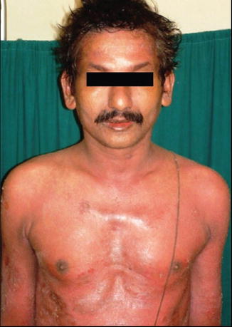
Erythroderma begins as erythematous and pruritic patches distributed throughout the body, which progressively or rapidly coalesce to involve 90 % or more of the cutaneous surface. The skin of these patients appears shiny, which is indicative of extensive dermal edema, and is typically bright red, suggesting widespread dilatation of dermal capillaries. The patient may complain of dryness and the feeling of having tight skin. The patient may also experience severe pruritus. Further physical examination typically reveals that the skin is warm to touch, with diffuse scaling throughout the body. Scaly patches may be present on the face, scalp, trunk, arms and legs, as well as the palms and soles. In some cases, the “nose sign” may be present, which occurs when the nasal and paranasal regions are not affected. However, this is not a diagnostic sign. Patients with drug eruption-related erythroderma may demonstrate evidence of leukonychia. Prolonged erythroderma may result in permanent hair loss throughout the body (alopecia) severe nail dystrophic changes, and coarse induration of the skin. Dark-skinned individuals may also exhibit widespread hypopigmentation.
Since the disease is commonly associated with, or precipitated by, an underlying cutaneous disorder, the above signs and symptoms may occur in confluence with evidence of the other disease. For example, patients with erythroderma associated with pre-existing psoriasis will experience the above, as well as psoriatic plaques (Fig. 23.1). Gotron’s papules, muscular weakness, and the classic heliotropic rash may be seen in patients with underlying dermatomyositis. Some patients may experience erythroderma in the context of the drug rash with eosinophilia and systemic symptoms (DRESS) syndrome (Fig. 23.2). The “red man syndrome” can present as a result of rapid intravenous infusion of antibiotics, particularly vancomycin, and consists of the acute appearance of an intensely erythematous rash that presents as erythroderma (Fig. 23.3). The erythema in red man syndrome tends to be continuous from the point of infusion, which is different than the appearance of the disease in other conditions, although this is certainly not a specific sign.


