Cellular Neurothekeoma Cellular neurothekeoma is a benign tumor of uncertain histogenesis that typically presents as a multilobular tumor centered in the dermis.
Nests and Aggregates of Tumor Cells Cellular neurothekeoma is generally characterized by multiple nests or aggregates of histiocytoid tumor cells divided by intervening fibrous bands.
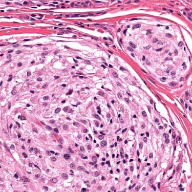
Bland Histiocytoid Cells in Cellular Neurothekeoma The nests are composed of histiocytoid cells with abundant pale cytoplasm and uniform-appearing, round nuclei.
Atypia and Mitotic Activity in Cellular Neurothekeoma Nuclear atypia and pleomorphism are seen in 25% of cellular neurothekeoma and do not indicate aggressive behavior. Mitotic activity is common and may even be brisk.
TERMINOLOGY
Definitions
• Rare dermal tumor of uncertain histogenesis composed of epithelioid cells in nests divided by fibrous septa
• Unrelated to dermal nerve sheath myxoma (conventional neurothekeoma)
CLINICAL ISSUES
Site
• Head/neck & upper extremity (most on face & shoulder)
Presentation
• Children & young adults (mean: 25 years; most < 40 years)
• Nearly 2:1 female predominance
• Painless solitary skin nodule, often present > 1 year
Rarely agminated (cluster of multiple lesions in 1 site)
Treatment
• Simple but complete excision
Prognosis
• Benign (even if atypical histologic features)
• Small subset recur, especially if incompletely excised
MACROSCOPIC
General Features
• Rounded or dome-shaped skin lesion, usually > 2 cm
MICROSCOPIC
Histologic Features
• Micronodular or lobulated dermal tumor
No epidermal involvement
Usually has infiltrative borders
50% also involve subcutis
• Epithelioid to spindled cells with abundant pale, eosinophilic cytoplasm, arranged in nests divided by dense fibrous septa
• Plexiform or focally sheet-like growth pattern sometimes
• Myxoid change common (30%)
Spindling more prominent in myxoid areas; may resemble pattern of dermal nerve sheath myxoma
• Multinucleated giant cells (osteoclastic or Touton) in some cases
• Nuclear atypia/pleomorphism in 25%
Atypical multinucleated tumor giant cells may be seen


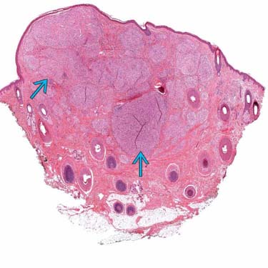
 centered in the dermis.
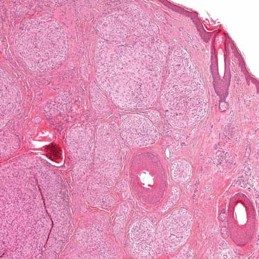
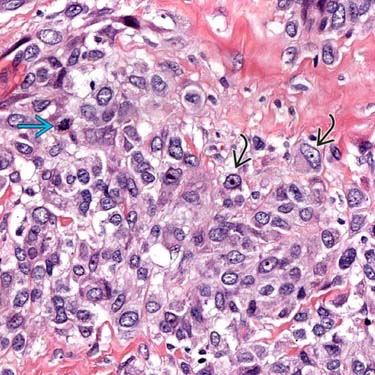
 and pleomorphism are seen in 25% of cellular neurothekeoma and do not indicate aggressive behavior. Mitotic activity
  is common and may even be brisk.


