16
Axilla and Brachial Plexus
The axilla (armpit) is the concave area on the inferior surface of the junction of the arm with the trunk. It has a base and four walls:
- The base is formed by hairy skin, subcutaneous fat, and axillary fascia.
- The apex is bounded by the posterior border of the clavicle, superior border of the scapula, and rib I.
- The anterior wall (anterior axillary fold) is formed by the pectoralis muscles (major and minor).
- The posterior border (posterior axillary fold) is formed by lateral parts of the latissimus dorsi, teres major, and subscapularis muscles.
- The medial wall consists of ribs I to IV and the intercostal and serratus anterior muscles.
- The lateral anterior wall is the intertubercular groove of the humerus.
- The apex is bounded by the posterior border of the clavicle, superior border of the scapula, and rib I.
Structural support of the axilla is provided by the lateral rib cage, clavicle, and scapula.
Nerves
The brachial plexus is a branching network of nerves that originates from the anterior primary rami of spinal nerves C5 to T1 (Fig. 16.1). It is enclosed with the axillary artery and vein within the axillary sheath, which is a prolongation of the prevertebral layer of cervical fascia that extends inferiorly behind the clavicle and into the axilla. The brachial plexus is composed of nerve roots, trunks, divisions, cords, and branches (Table 16.1).
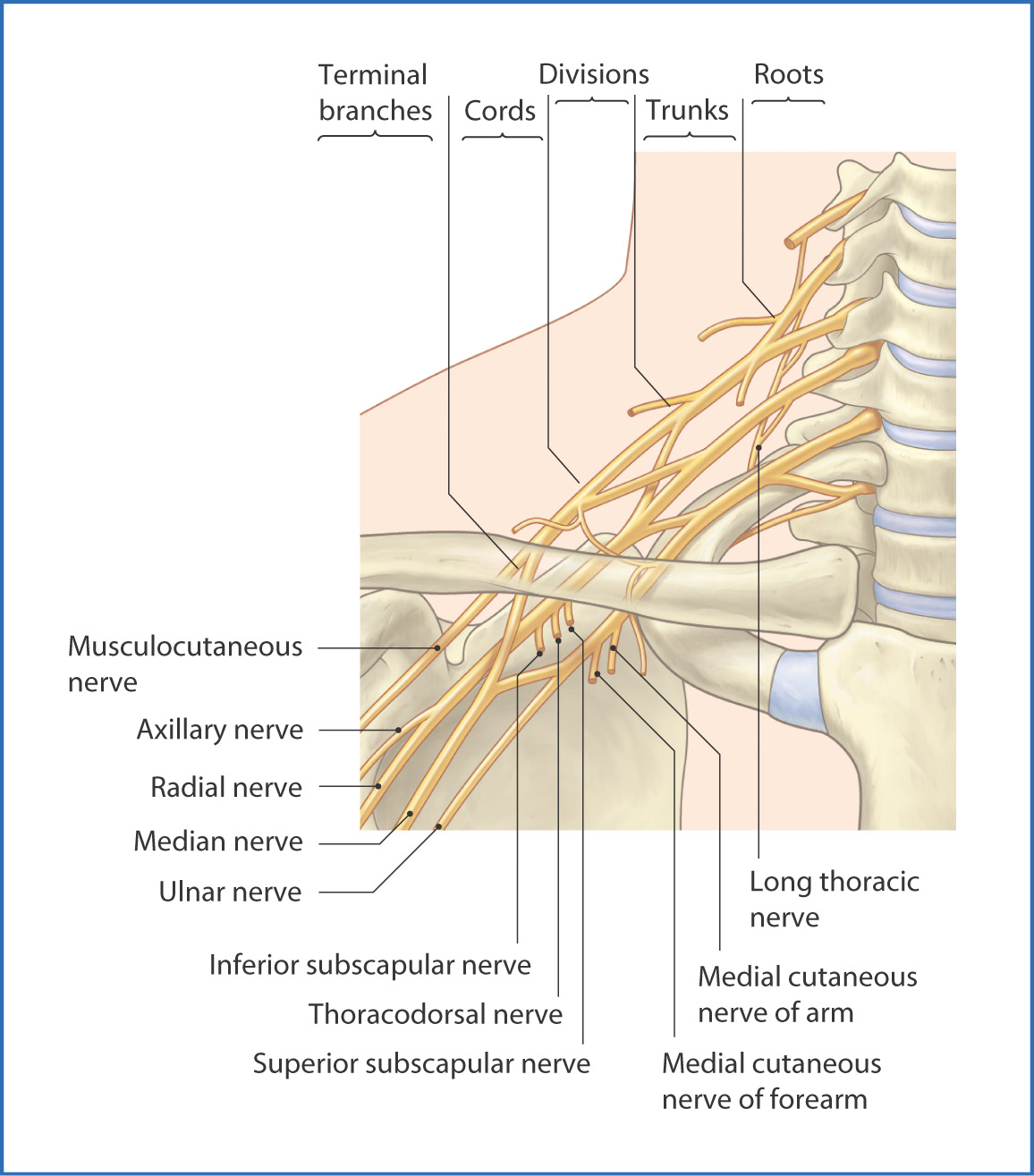
FIGURE 16.1 Brachial plexus.
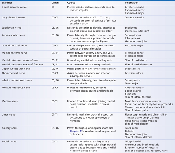
TABLE 16.1 Branches of the Brachial Plexus
The nerve roots emerge from the spinal canal through the intervertebral foramina of the lower cervical vertebrae. They are situated in the deep posterior region of the neck (see Chapter 13) between the anterior scalene and middle scalene muscles just superior to rib I. Two nerves come off the spinal roots, the dorsal scapular nerve (to the levator scapulae, rhomboid major, and rhomboid minor muscles) and the long thoracic nerve (to the serratus anterior muscle).
As they pass laterally, the spinal roots unite to form three trunks (superior, middle, and inferior trunks). The superior trunk is formed by the upper two roots (C5, C6), the middle trunk by C7, and the inferior trunk by C8 and T1. Two nerves leave the superior trunk—the suprascapular nerve and subclavian nerve.
Behind the clavicle, each trunk divides into anterior and posterior divisions. Nerves do not usually leave the divisions of the brachial plexus. The anterior division of the superior and middle trunks forms the lateral cord, the anterior division of the inferior trunk continues as the medial cord, and the posterior divisions of all three trunks form the posterior cord. These cords are named according to their relationship to the second part of the axillary artery. Several nerves leave the cords:
- The lateral pectoral arises from the lateral cord and becomes the musculocutaneous nerve.
- The medial pectoral nerve, medial cutaneous nerve of the arm, and medial cutaneous nerve of the forearm arise from the medial cord.
- The superior subscapular, thoracodorsal, and inferior subscapular nerves arise from the posterior cord.
- The medial pectoral nerve, medial cutaneous nerve of the arm, and medial cutaneous nerve of the forearm arise from the medial cord.
Five terminal branches (nerves) of the brachial plexus arise from the three cords. Each cord divides into medial and lateral branches:
- The lateral branch of the posterior cord becomes the axillary nerve (C5 to C6).
- The medial branch of the posterior cord becomes the radial nerve (C5 to T1).
- The lateral branch of the lateral cord becomes the musculocutaneous nerve (C5 to C7).
- The medial branch of the lateral cord is joined by the lateral branch of the medial cord to form the median nerve (C6 to T1).
- The medial branch of the medial cord becomes the ulnar nerve (C7 to T1).
- The medial branch of the posterior cord becomes the radial nerve (C5 to T1).
Arteries
Branches of the axillary artery (Fig. 16.2) supply blood to the structures of the axilla. The axillary artery, a continuation of the subclavian artery, arises at the lateral border of rib I inferior to the roots of the brachial plexus and between the anterior and middle scalene muscles. It continues into the axilla and terminates at the lower border of the teres major muscle, where it becomes the brachial artery. The axillary artery is divided into three parts based on its relationship to the pectoralis minor muscle:
Stay updated, free articles. Join our Telegram channel

Full access? Get Clinical Tree


