21
Anterior Forearm
The forearm is the part of the upper limb between the elbow and wrist. It is divided into anterior and posterior compartments by the interosseous membrane, which joins the radius and ulna. The anterior compartment of the forearm contains several muscles that flex the wrist and digits (fingers). The forearm is supported by the radius and ulna.
The superior part of the ulna bears the coronoid process and olecranon, which articulate with the humerus at the elbow joint. Distally, the ulna is narrow; its small head articulates with the radius through the distal radio-ulnar joint and indirectly with carpal (wrist) bones via an interarticular disc.
The radius is shorter than the ulna and is lateral in the forearm. Its distal end is large and articulates directly with the carpal bones at the radiocarpal joint. Proximally, the anular ligament of the radius binds the head of the radius to the ulna and allows the rotation of the forearm necessary for pronation and supination. The arrangement of the radius and ulna is unique in that weight is transferred from the wrist to the radius and then to the humerus via the ulna.
Muscles
The muscles in the anterior compartment of the forearm (Figs. 21.1 and 21.2) can be divided into a superficial group of five muscles and a deep group of three muscles. From lateral to medial, the superficial group comprises the
- pronator teres
- flexor carpi radialis
- palmaris longus
- flexor carpi ulnaris
- flexor digitorum superficialis
- flexor carpi radialis
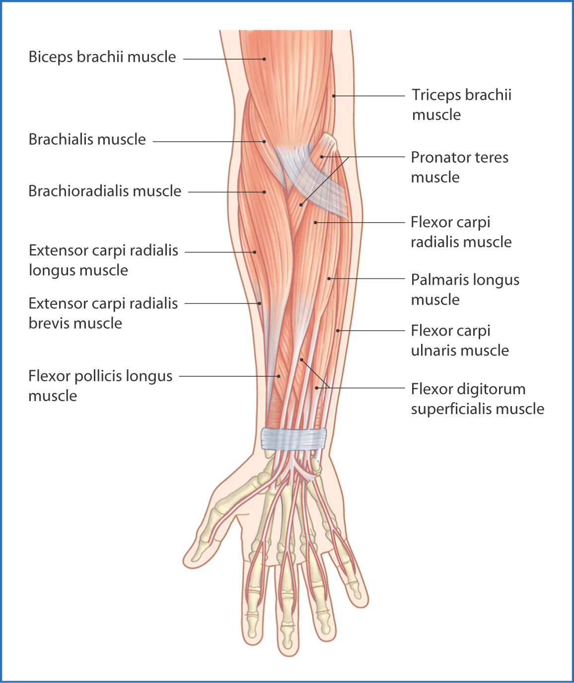
FIGURE 21.1 Superficial muscles of the anterior aspect of the right forearm.
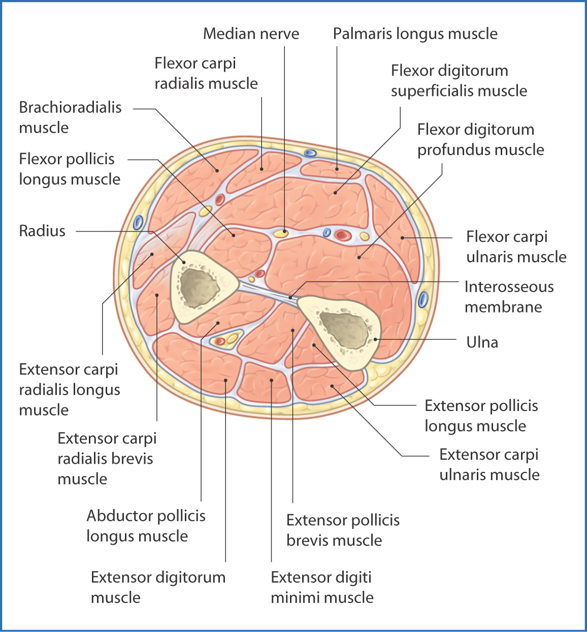
FIGURE 21.2 Cross section through the right forearm (viewed from distal to proximal).
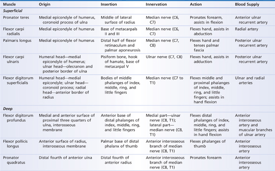
The superficial muscles have a similar origin at the common flexor tendon, which originates from the front of the medial epicondyle of the humerus. The flexor digitorum superficialis muscle is unique in that it has three heads of origin (Table 21.1) and sends four tendons to the middle phalanx of the index, middle, ring, and little fingers. The flexor digitorum superficialis and other superficial muscles flex the hand and fingers.
TABLE 21.1 Anterior Forearm Muscles
The deep muscles in the anterior compartment of the forearm are the
The flexor digitorum profundus muscle arises from the upper three quarters of the medial and anterior surface of the ulna and the interosseous membrane (Fig. 21.3). It inserts onto the base of the distal phalanges of the medial four digits via four tendons.
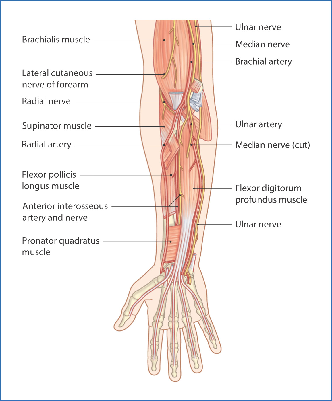
FIGURE 21.3 Deep structures of the anterior aspect of the right forearm.
The flexor pollicis longus muscle arises from the upper two thirds of the anterior surface of the radius and inserts into the distal phalanx of the thumb. The flexor digitorum profundus and flexor pollicis longus muscles flex the digits and assist in flexion of the hand.
The pronator quadratus muscle takes origin from the distal anterior surface of the ulna and inserts onto the medial and anterior surfaces of the distal end of the radius. With this unique layout it acts to pronate the forearm.
Nerves
The median nerve (C6 to T1) provides the main innervation of the anterior aspect of the forearm in that it innervates  of the 8 muscles. It originates from the brachial plexus, descends in the arm, and passes into the cubital fossa where it supplies the pronator teres, flexor carpi radialis, and palmaris longus muscles. In the cubital fossa the median nerve pierces the pronator teres muscle, and on leaving this muscle it branches to give off the anterior interosseous nerve
Stay updated, free articles. Join our Telegram channel

Full access? Get Clinical Tree


