Grade 3 tumors: Poorly differentiated neuroendocrine carcinoma (small or large cell type)
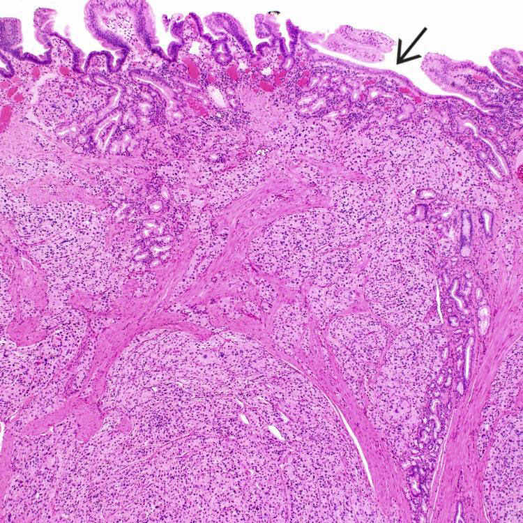
Low-power view shows a well-differentiated neuroendocrine neoplasm infiltrating the ampullary region. The overlying mucosa is attenuated and shows gastric foveolar metaplasia
 .
.
Tumor cells are typically uniform with round nuclei, salt and pepper chromatin, inconspicuous nuclei, and moderate amounts of eosinophilic cytoplasm. Focal glandular formation
 is noted. Mitotic figures are difficult to find.
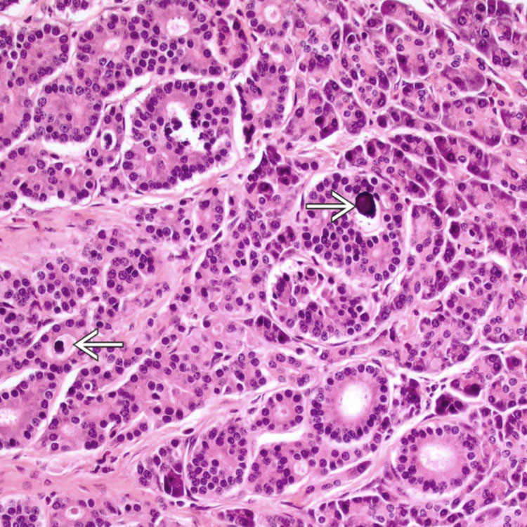
Well-differentiated neuroendocrine neoplasm of the ampulla shows extensive glandular formation. Psammomatous calcifications are noted in the lumina of some of the glands
 . These features are more commonly seen in somatostatin-producing tumors.
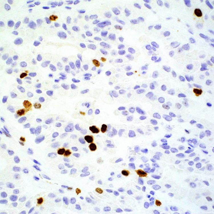
Well-differentiated neuroendocrine neoplasm of the ampulla shows positive immunostain for Ki-67 in ∼ 5% of tumor cells. The tumor is thus graded as intermediate (grade 2), even though the mitotic rate is < 2 per 10 HPF.


