Neonatal Hemochromatosis

 Liver


image Pancreas

image Heart

image Thyroid

image Minor salivary glands


• Abnormal iron studies

• Liver and multiorgan failure


Macroscopic


• Shrunken liver

• Cirrhosis


Microscopic


• Marked lobular necrosis with collapse

• Regenerative nodules

• Pseudoacinar formation

• Intracanalicular bile plugs

• Giant hepatocytes

• Iron deposition in hepatocytes and ductules as well as in organs outside of liver


Top Differential Diagnoses


• Virus infection
image CMV

image Echovirus

image Herpes simplex virus

• Neonatal lupus

• Tyrosinemia

image
Lobular Necrosis
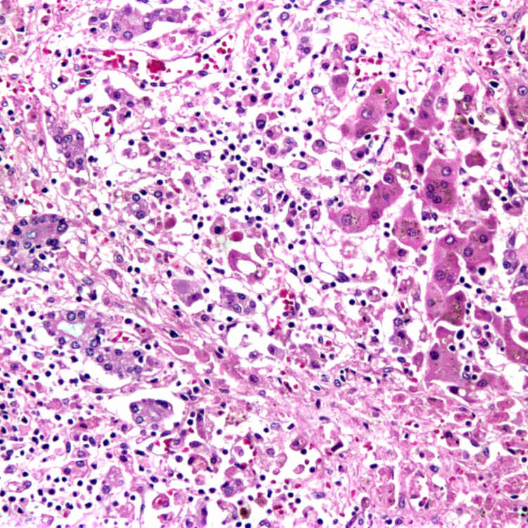
H&E in this case of neonatal hemochromatosis shows lobular necrosis and collapse of the hepatocellular cords with residual hepatocytes and bile ductules.


image
Perl Iron Stain
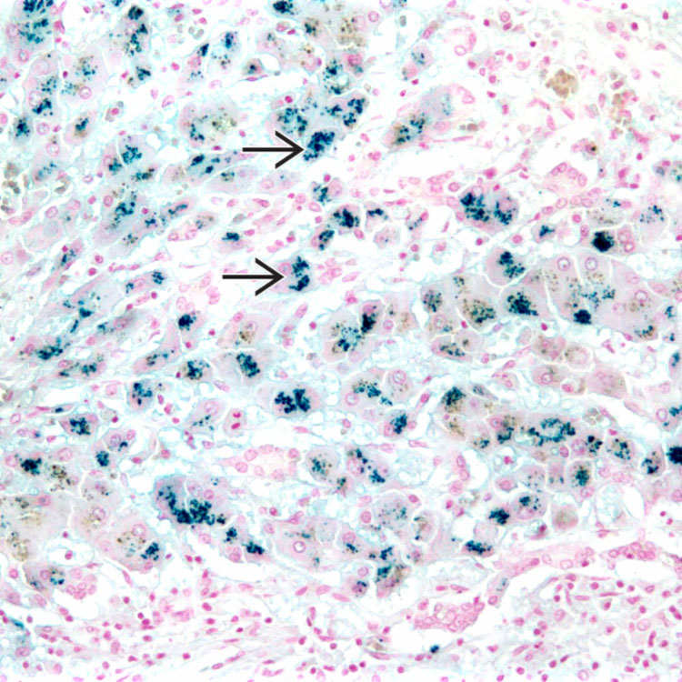
Perl iron stain in this case of neonatal hemochromatosis shows marked iron deposition image within the hepatocytes.

image
Submassive Hepatocellular Necrosis
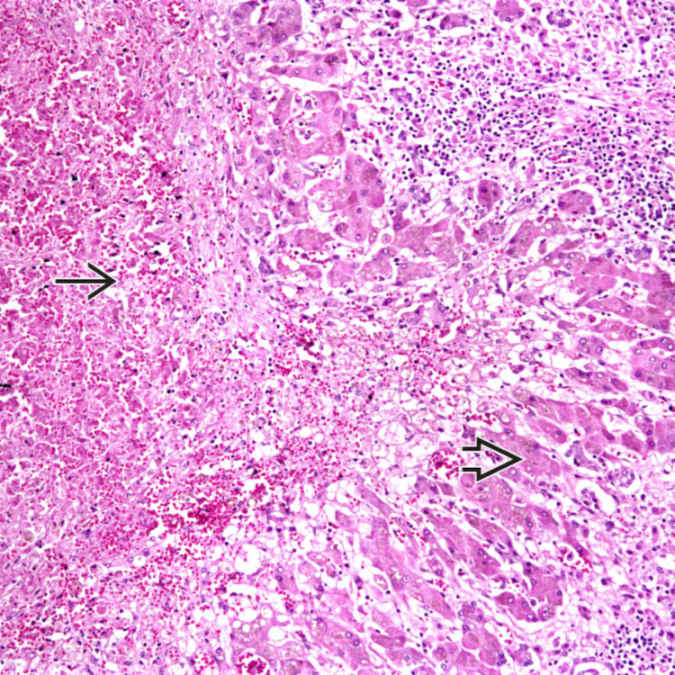
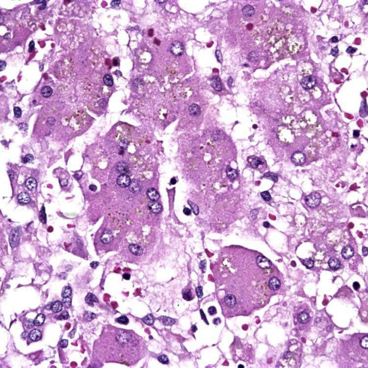
Higher power of this case of neonatal hemochromatosis shows submassive hepatocellular necrosis image with a rim of residual hepatocytes image .

image
Giant Cells
Giant cells are seen in this case of neonatal hemochromatosis.


TERMINOLOGY


Abbreviations


• Neonatal hemochromatosis (NH)


Synonyms


• Congenital hemochromatosis

• Neonatal iron storage disease

• Gestational alloimmune disease


Definitions


• Severe liver disease with iron overload in liver and other organs (distribution similar to hereditary hemochromatosis)
image Fetal or perinatal onset

Only gold members can continue reading. Log In or Register to continue

Stay updated, free articles. Join our Telegram channel

Apr 20, 2017 | Posted by in PATHOLOGY & LABORATORY MEDICINE | Comments Off on Neonatal Hemochromatosis

Full access? Get Clinical Tree

Get Clinical Tree app for offline access