Congenital Hepatic Fibrosis

 Depending on severity of portal hypertension, biliary infection, and renal disease


Microscopic


• Marked portal expansion by fibrous tissue
image Portal-to-portal bridging fibrosis with broad septa

• Increased number of irregularly shaped, ectatic, branching, and anastomosing bile ducts
image Sometimes line border of portal tracts or fibrous septa in ductal plate configuration

image Inspissated bile may be seen in lumina of ducts

• Portal vein branches may be hypoplastic or reduced in number, or show cavernous transformation

• Hepatic artery branches may be hypertrophic or abnormally numerous


Top Differential Diagnoses


• Cirrhosis

• von Meyenburg complexes

image
Gross Appearance
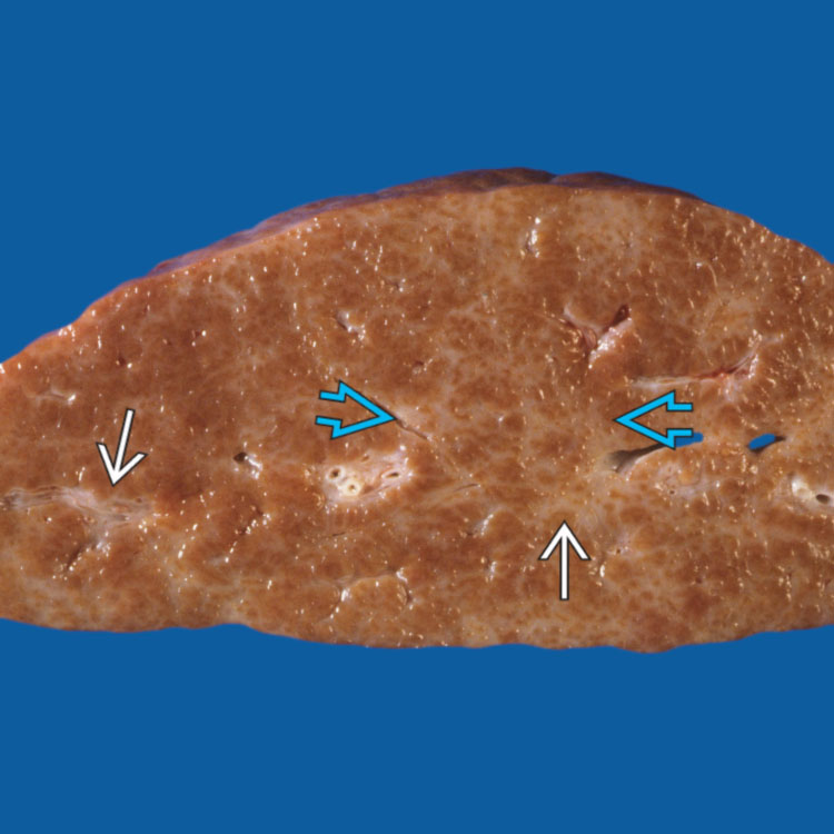
A gross photograph of congenital hepatic fibrosis shows white fibrous bands image that divide the liver parenchyma in a reticular pattern image. There is no definite nodularity, as opposed to cirrhosis.


image
Bridging Fibrosis and Aberrant Ducts
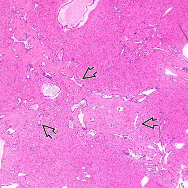
Marked fibrous portal expansion with bridging fibrosis and numerous ectatic and irregularly shaped bile ducts are present, some of which are characteristically located at the interface with liver parenchyma image. Note that there is no significant inflammation in this case.

image
Inspissated Bile
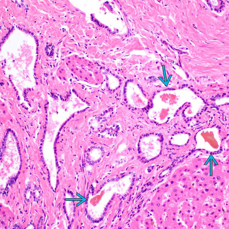
The aberrantly formed bile ducts are lined by cuboidal biliary epithelium, and some contain inspissated bile image. There is no significant inflammation.

image
Vascular Anomalies
Portal vein branches may be hypoplastic or reduced in number. This fibrotic portal tract contains multiple prominent hepatic artery branches image
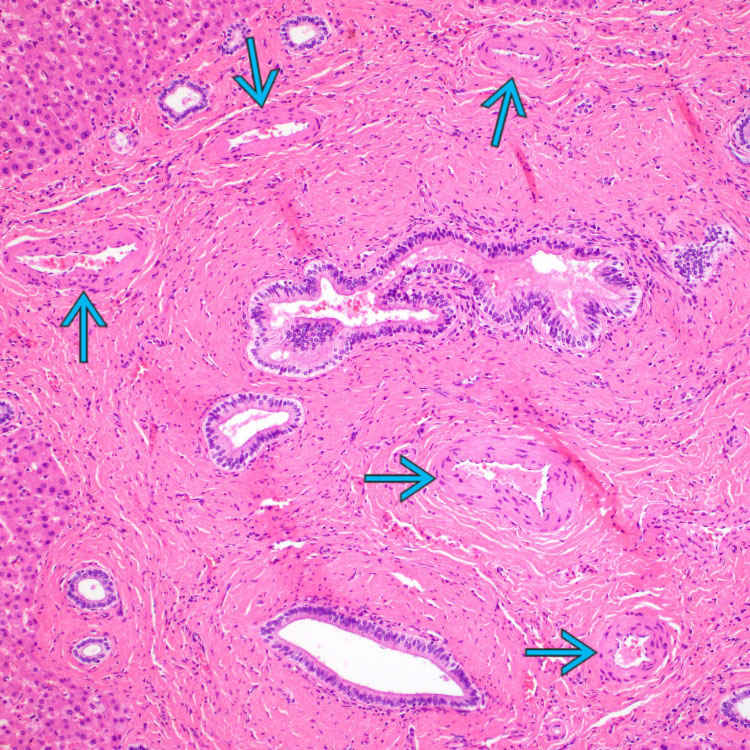
, but portal veins are not recognizable.


TERMINOLOGY


Abbreviations


• Congenital hepatic fibrosis (CHF)


Definitions


• Variant of ductal plate malformation that leads to portal and bridging fibrosis, proliferation of aberrant duct profiles, and portal hypertension


ETIOLOGY/PATHOGENESIS


Developmental Anomaly


• Ductal plate malformation at level of interlobular bile ducts

• Persistence of excess embryonic bile ducts

• Primarily autosomal recessive inheritance, rarely autosomal dominant


Disease Associations


• Autosomal recessive polycystic kidney disease
image Most common coexisting condition

image Affecting 1 in 20,000 live births

image Mutations in PKHD1 gene encoding fibrocystin/polyductin located in primary cilia

– Most common ciliopathy of childhood

Only gold members can continue reading. Log In or Register to continue

Stay updated, free articles. Join our Telegram channel

Apr 20, 2017 | Posted by in PATHOLOGY & LABORATORY MEDICINE | Comments Off on Congenital Hepatic Fibrosis

Full access? Get Clinical Tree

Get Clinical Tree app for offline access