Form of noncirrhotic portal hypertension


Microscopic
• Abnormal hepatic architecture with lack of consistent relationship between portal tracts and central veins
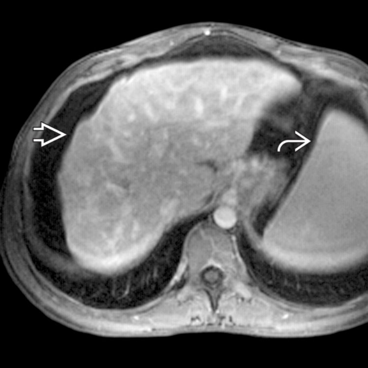
Abdominal MR shows an irregular hepatic surface with a shrunken appearance
 and heterogeneous hepatic parenchyma due to portal fibrosis. Note the presence of splenomegaly
  .
.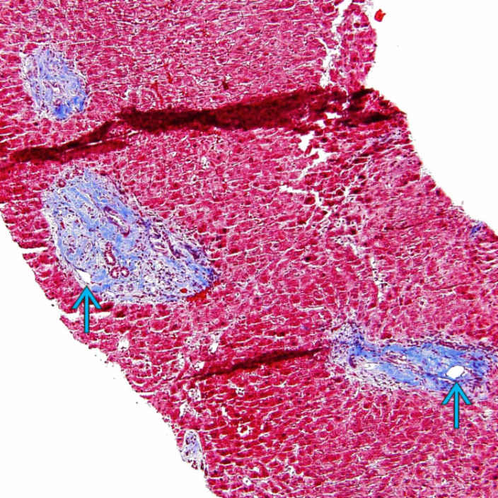
Trichrome stain highlights abnormal approximation of portal tracts seen in a needle biopsy from a patient with portal hypertension. Portal fibrosis and narrowed portal veins
 are evident. No central veins are seen between portal tracts in this field. Note the absence of cirrhosis.
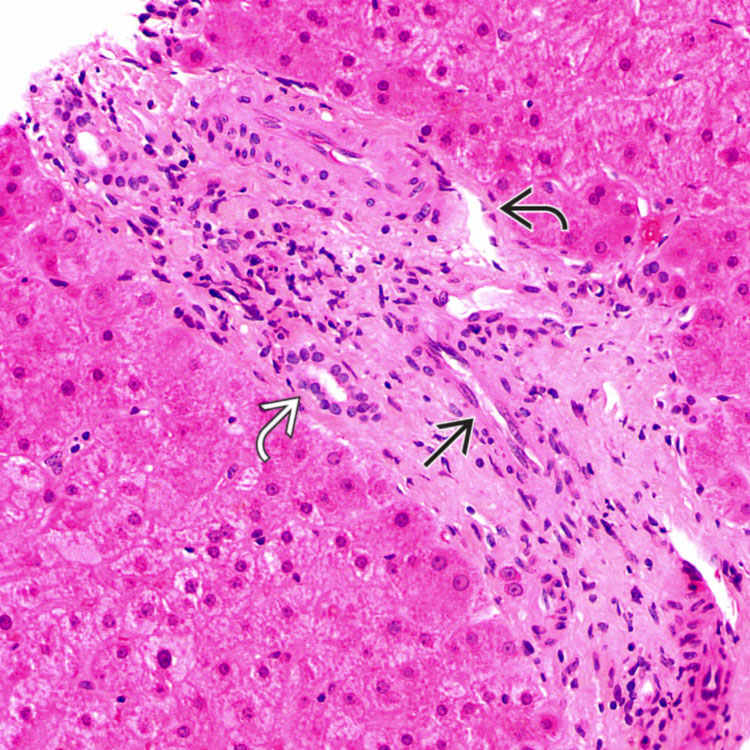
This portal tract is expanded by fibrosis, with marked narrowing of the portal vein
 . Note the presence of normal-caliber hepatic artery
  and bile duct
  . There are no significant inflammatory cell infiltrates in the portal tract.
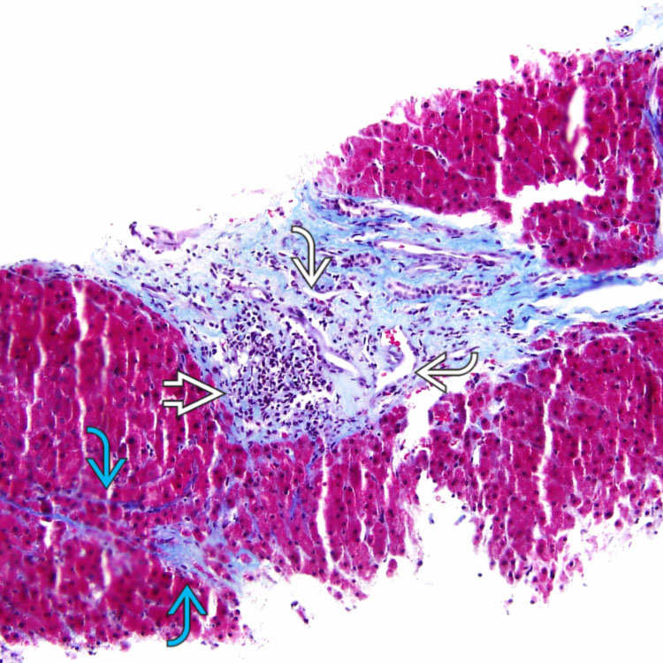
 , and multiple slit-like spaces
  representing narrowed portal veins. Focal perisinusoidal collagen deposition
  is also noted in this biopsy.


