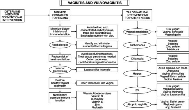
• Symptoms could overlie serious problem (e.g., chronic cervicitis or sexually transmitted disease). If infectious, agent may ascend the genital tract, leading to endometritis, salpingitis, and pelvic inflammatory disease (PID), leading to tubal scarring, infertility, or ectopic pregnancies. • Implicated in recurrent UTIs by acting as reservoir of infectious agents. • Some vaginal infections during pregnancy increase the risk of miscarriage and, if present at delivery, cause neonatal in-fections. • Some forms of vaginitis are linked to cervical cellular abnormalities and increased risk of cervical dysplasia. span id=”pgbrk757″ epub:type=”pagebreak” title=”757″/> Hormonal, irritant, and infectious; each is divided into subgroups based on etiology. — Atrophic vaginitis: affects perimenopausal and postmenopausal women. The structure and function of vaginal tissues undergo change with declining estrogenic stimulation, including estrogen loss after oophorectomy. Vaginal epithelium atrophies from a lack of estrogenic stimulation; this may cause a decrease in Lactobacillus in the vagina, more alkaline pH, vaginal thinning, less lubrication, more easily irritated and inflamed tissue, adhesions, dyspareunia, and increased susceptibility to infection. Most common symptoms are itching or burning and thin, watery discharge, occasionally blood tinged (any vaginal bleeding in postmenopausal women requires complete workup to rule out endometrial hyperplasia and endometrial carcinoma; see baseline evaluation in the chapter on menopause). — Increased vaginal discharge: increased normal secretions in the absence of other symptoms. Diagnosis of physiologic vaginitis often is applied but is inappropriate because no inflammation exists. Increased discharge often reflects increased hormonal stimulation (pregnancy or some stages of menstrual cycle). This is primarily a diagnosis of exclusion after ruling out other causes. Usually no further treatment is required other than reassurance. Overly zealous douching or washing briefly alleviates symptoms but may aggravate the condition by causing irritant vaginitis. — Caused by physical or chemical agents damaging delicate vaginal membranes. Identified by careful history and examination. — Chemical vaginitis: medications or hygiene products can irritate vaginal mucosa. Allergic vaginitis is damage elicited by immunologic reaction to a product rather than direct toxic reaction. Perfumed toilet paper, douches, spermicides, condoms, and lubricants can be irritating agents. — Traumatic vaginitis: injury caused by physical agents or sexual activity. — Foreign body vaginitis: foul discharge may indicate foreign body in vagina. Most common are forgotten tampons, contraceptive devices, and pessaries. Very young girls and adolescents may leave foreign bodies in their vaginas from exploring their bodies; they either forget or are too embarrassed to tell someone that they cannot retrieve the item. Also, semen may be an irritant in some women. — Ninety percent of vaginitis in reproductive-aged women is caused by bacterial vaginosis (BV), candidiasis, or trichomoniasis. BV is most common. Less-common causes include herpes simplex virus, gonorrhea (GC), and chlamydia. Infections of the vulva can cause local itching and/or discharge: folliculitis, hydradenitis, scabies, condyloma, herpes, syphilis, candida. Rare conditions of vulva causing itching and/or discharge include chancroid, lymphogranuloma venereum inguinale, and molluscum contagiosum. — May be sexually transmitted or a result of a disturbance of the delicate ecosystem of the healthy vagina. It often involves common organisms found in the cervix and vagina of healthy, asymptomatic women. — Unifying factor in pathogenesis of pelvic infections is not which microbes are present, but rather the cause of patient susceptibility. — Factors influencing vaginal environment are pH, glycogen content, glucose level, presence of other microbes (lactobacilli), natural flushing action of vaginal secretions, presence of blood, and presence of antibodies and other compounds in vaginal secretions; all are affected by a woman’s internal milieu and general health. — Immune dysfunction predisposes increased vaginal infections: nutritional deficiencies, medicines (e.g., steroids), pregnancy, or serious illness. — Other predisposing factors are diabetes mellitus, hypothyroidism, leukemia, Addison’s disease, Cushing’s syndrome, pregnancy, and Candida infections. — Predisposing factors for sexually transmitted disease: increased number of sexual partners, unusual sexual practices, type of birth control (barriers reduce risk), unsafe sex, birth control pills, steroids, antibiotics, tight-fitting garments, occlusive materials, douches, chlorinated pools, perfumed toilet paper, and decrease in Lactobacillus in vagina. • Trichomonas vaginalis: flagellated protozoan found in the lower genitourinary tract of men and women. Human beings are the only host. Sexual intercourse is the primary mode of dissemination. Trichomonas do not invade tissues and rarely cause serious complications. — Most frequent symptoms are leukorrhea, itching, and burning. Discharge is malodorous, greenish yellow, and frothy. — “Strawberry cervix” with punctate hemorrhages is found only in a small percentage of Trichomonas cases. — Grows optimally at pH = of 5.5-5.8. Conditions elevating pH (e.g., increased progesterone) favor Trichomonas growth. Vaginal pH of 4.5 in women with vaginitis suggests agent other than Trichomonas. — Saline wet mount of fresh vaginal fluid shows small motile organisms, confirming diagnosis in 80%-90% of symptomatic carriers. • Candida albicans: 2.5-fold increase in candidal vaginitis has occurred in the past 20 years, paralleling the declining incidence of GC and Trichomonas. Contributing factors are increased use of antibiotics, which changes the vaginal ecosystem to favor Candida. — One hundred percent correlation between genital and gastrointestinal Candida cultures: significant intestinal colonization with Candida may be the single most significant predisposing factor in vulvovaginal candidiasis. However, this has not been confirmed by adequate research. Steroids, oral contraceptives, and diabetes mellitus contribute. Candidiasis is 10-20 times more frequent during pregnancy because of elevated vaginal pH, increased vaginal epithelial glycogen, elevated blood glucose, and intermittent glycosuria. — Candidiasis is three times more prevalent in women wearing pantyhose than in those wearing cotton underwear; pantyhose prevents drying of area. — Four or more episodes of vulvovaginal candidiasis in 1 year classify the patient as having recurrent disease. Cause may be non-albicans strains of Candida—resistant strains generated by antifungals. Three main theories of why women get recurring yeast vaginitis: (1) intestinal reservoir migrating into vagina, (2) sexual partner is source of recurrence, and (3) vaginal relapse forms small residual numbers of yeast after treatment. Research supports this last theory. Women with recurrent infections have an abnormal immune response, increasing susceptibility. — Allergies can cause recurrent candidiasis, which resolves after allergies treated. — Primary symptom of candidiasis is vulvar itching (sometimes severe). Burning of vulva, exacerbated by urination or vaginal sexual activity, also occurs. Thick, curdy or “cottage cheese” discharge that adheres to vaginal walls may be present. Such discharge is strong evidence of yeast infection, but its absence does not rule out Candida. Less than 20% of symptomatic candidiasis displays classic thrush patches. In addition, an increase or change in the consistency of the vaginal discharge is common. — Other clues include erythema of vulva and excoriations from scratching. Vaginal pH is not usually altered. — Neither the character of discharge nor the symptoms are sufficient alone for diagnosis of Candida. Wet mount in saline or 10% KOH; mycelia are confirmatory. Budding forms of yeast are found in normal and symptomatic vaginas, but mycelial stage is only found in symptomatic women. • Nonspecific vaginitis (NSV): vaginitis not caused by Trichomonas, GC, or Candida. • Bacterial vaginosis (BV): shift in flora predominance from lactobacilli to anaerobes and facultative bacteria that degrade mucins of natural gel barrier on vaginal epithelium, causing characteristic vaginal discharge. Destruction of mucins exposes cervical epithelium and allows other organisms to affect cervix, resulting in appearance of clue cells. Epithelial surface disruption causes immunologic shifts: upregulation of interleukin-1-beta and decrease in protectant molecules (e.g., secretory leukocyte protease inhibitor). — Three factors shift dominance from lactobacilli to anaerobes and facultative bacteria: sexual activity, douching, and absence of peroxide-producing lactobacilli. A new sexual partner or frequent sexual activity increases incidence of BV. Routine douching is linked to loss of vaginal lactobacilli and occurrence of BV. Women who regularly douche for hygiene purposes have BV twice as often as women who do not routinely douche. Women lacking vaginal peroxide–producing lactobacilli may not have had normal lactobacilli at menarche, or lactobacilli were eliminated by broad-spectrum antibiotics. — Other factors increasing risk for BV: cigarette smoking suppresses immune response. Racial background: Hispanic women have a 50% greater risk and African-American women have twice the risk. African-American women practice douching twice as often as white women; African-American women also are less likely to have vaginal lactobacilli. — Examination for BV: medical history; observe discharge to determine vaginal pH. Symptomatic BV releases fishy odor. Discharge is thin, dark, or dull grey. Itching is uncommon in BV unless discharge is profuse. Perform whiff test and wet mount to detect clue cells and observe vaginal flora. Abundance of various bacteria and absence or decrease of lactobacilli suggests BV. Numbers of bacteria increase from 100-fold to 1000-fold. Diagnosis of BV requires three of four criteria: 1. Thin, dark, or dull grey, homogeneous, malodorous discharge that adheres to vaginal walls 3. Positive KOH (whiff/amine test) 4. Clue cells on wet mount microscopic exam. pH is elevated to 5.0-5.5; correlation exists between elevated pH and presence of odor.
Vaginitis and Vulvovaginitis
GENERAL CONSIDERATIONS

TYPES OF VAGINITIS
![]()
Stay updated, free articles. Join our Telegram channel

Full access? Get Clinical Tree


Vaginitis and Vulvovaginitis
