and John E. Skandalakis1
(1)
Centers for Surgical Anatomy and Technique, Emory University School of Medicine Piedmont Hospital, Atlanta, GA, USA
Abstract
The role of ligaments as support for the uterus, fallopian tubes, ovaries, and bladder is described. The ovary has the capacity to produce many benign and malignant tumors; maximal effort should be focused on the preservation of useful function when the presumed histology is benign.
Both the surgical technique of abdominal hysterectomy and the significance of the procedure to the patient are covered. Different incisions are preferred for cancer, tubo-ovarian abscess, limited-dimension benign disease, and large fibroids or extensive endometriosis. A step-by-step general technique of hysterectomy, suitable when there is no significant disruption of the normal anatomy, is proposed; modifications are needed according to disease processes encountered. A modification for bilateral salpingo-oophorectomy is provided. A new section on ovarian cystectomy discusses treatment of ruptured and unruptured cysts in premenopausal and postmenopausal patients.
Anatomy
Relations and Positions of the Uterus, Tubes, and Ovaries
The uterus lies in the pelvis between the bladder and rectum. The normal position of the uterus—whether anteverted, midplane, or retroverted—is maintained by the round ligaments. The ligaments insert laterally, anterior to the fallopian tubes, and then plunge into the pelvic sidewall. The round ligaments may be viewed as the roof of the broad ligament. The broad ligament contains the blood supply, lymphatic channels, and nerves of the corpus uteri.
The ureter courses just below the insertion of the uterine artery into the lower uterine segment, and it should be identified clearly to avoid injuring it. The lower uterine segment and cervix are bordered anteriorly by the bladder and posteriorly by the rectum. Moving downward, the surgeon encounters the uterosacral ligaments, which provide critical support to the uterus. Laterally and downward the broad ligament joins the cardinal ligament until the cervical/vaginal junction is reached.
The fallopian tubes emerge from the fundus and are in close proximity to the ovaries. The mesosalpinx descends from the tubes. The ovaries are joined to the uterus via the utero-ovarian ligament and to the pelvic sidewall by the infundibulopelvic ligament. The infundibulopelvic ligament contains the ovarian vessels.
Vascular System of the Uterus, Tubes, and Ovaries
Arterial Supply
The uterine artery arises from the internal iliac artery, as do the cervical, vaginal, and other collateral vessels.
Venous Supply
The veins follow a course analogous to the internal iliac vein. The ovarian artery and vein course in a cephalad direction and have no pelvic origin.
Lymphatic Drainage
Coursing parallel to the internal iliac vessels, the drainage from the corpus uteri and cervix ends in the deep pelvic lymph nodes. The drainage from the ovaries is in a cephalad and midline direction, coursing to the periaortic nodes, adjacent to the inferior vena cava and aorta.
Technique
Abdominal Hysterectomy and Bilateral Salpingo-Oophorectomy
In addition to understanding the surgical technique of hysterectomy, the surgeon should realize the significance of the procedure to the patient. Removal of the uterus will, under usual circumstances, sterilize the patient. Removal of the ovaries will result in castration of the patient. The possible need for estrogen replacement therapy, with all its attendant controversies, might then develop. It is beyond the scope of this chapter to consider these issues, but the surgeon is encouraged, when possible, to clearly understand the patient’s wishes and perspective.
The incision for hysterectomy is often determined by the indications for the procedure. When cancer is suspected, a midline incision is performed from the umbilicus to the symphysis pubis. For large fibroids or extensive endometriosis, a Maylard incision (muscle-cutting transverse incision) might be performed. A tubo-ovarian abscess can be approached with either a Maylard or a midline skin incision. For benign disease of limited dimension, a Pfannenstiel incision (low transverse muscle-spreading incision) is often performed.
The following general technique of hysterectomy is suitable when there is no significant disruption of the normal anatomy; modifications are needed according to disease processes encountered.
PREOPERATIVE: Prepare the vagina carefully with a Betadine solution; administer antibiotics.
Get Clinical Tree app for offline access

Step 1. Make incision. Generally a transverse muscle-splitting or muscle-cutting incision is performed. This choice of incision is associated with less postoperative pain and a more desirable cosmetic result. For a transverse incision, the skin is cut approximately 2 cm above the symphysis pubis and 3–4 cm to each side of the midline.
Step 2. Incise the rectus abdominis fascia and extend this incision laterally. Grasp the superior edge and sharply dissect the underlying muscle away from the fascia with Metzenbaum scissors. Bleeding from perforated vessels can be controlled with electrocautery.
Step 3. Take a Kelly clamp and identify the midline of the muscle. Gently separate the bellies and extend the incision superiorly and inferiorly, reaching the limits of your previous dissection.
Step 4. Grasp a peritoneal fold. Enter cautiously to avoid injury to underlying bowel. Extend the peritoneal incision superiorly and inferiorly.
Step 5. If a neoplasm is suspected, take pelvic washings and submit for cytology. If an infectious process is apparent, take fluid for culture.
Step 6. Place Kelly clamps on each corner of the uterus encompassing the round ligaments, utero-ovarian ligaments, and fallopian tubes.
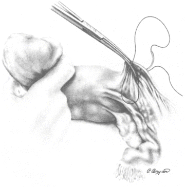
Step 7. Place gentle traction on the clamps. With a curved Heaney, Zeppelin, or Masterson pelvic clamp, clamp the round ligament approximately 2 cm lateral to its uterine insertion. Cut the round ligament with a scalpel, leaving a pedicle approximately 3 mm distal to the clamp. Secure the pedicle with a 0 or 2–0 Vicryl suture ligature. All subsequent sutures are the same unless otherwise indicated (Fig. 18.1).

Figure 18.1.
Round ligament is clamped, ligated, and cut. Broad ligament is opened (from HW Jones III. Hysterectomy. In: JA Rock and HW Jones III, eds. TeLinde’s Operative Gynecology, 9th ed. Philadelphia: Lippincott Williams & Wilkins, 2003, p. 810, with permission).
Stay updated, free articles. Join our Telegram channel

Full access? Get Clinical Tree


