and John E. Skandalakis1
(1)
Centers for Surgical Anatomy and Technique, Emory University School of Medicine Piedmont Hospital, Atlanta, GA, USA
Abstract
The surgical anatomy of the palm is complex. Patients with chronic carpal tunnel syndrome may benefit from a number of different nonsurgical and surgical treatments. Surgery is usually performed on an outpatient basis. During surgery, the carpal tunnel contents should be checked for anatomical abnormality, degenerative elements, and/or inflammatory conditions.
Anatomy
The surgical anatomy and anatomical entities related to the carpal tunnel syndrome are presented through illustrations and tables (Figs. 19.1, 19.2, 19.3, 19.4, and 19.5; Tables 19.1, 19.2, and 19.3).
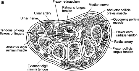
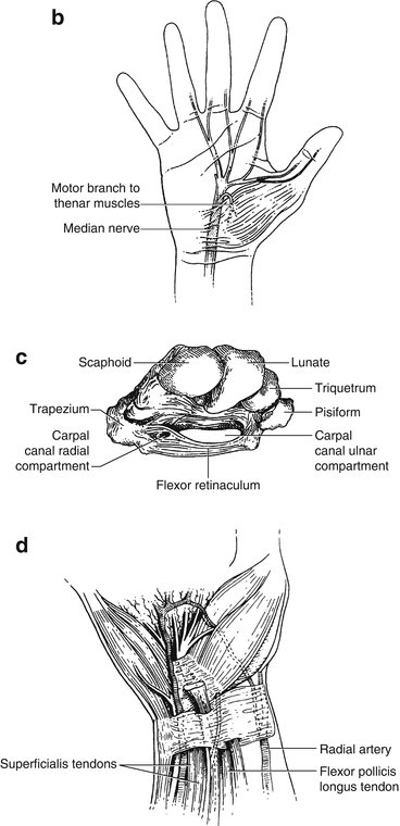
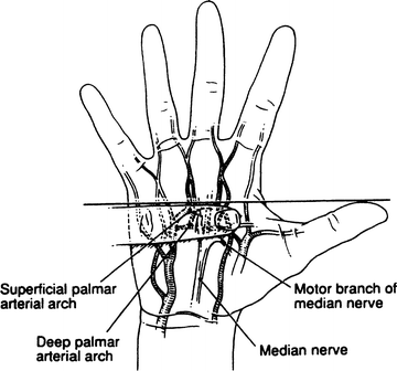
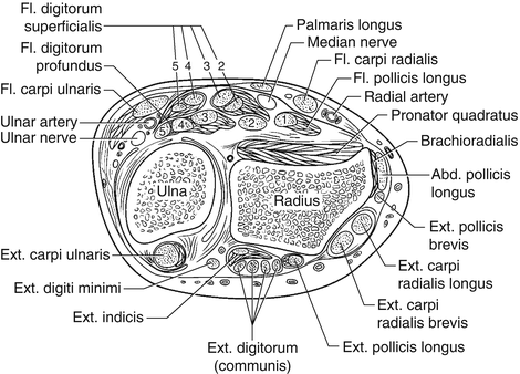
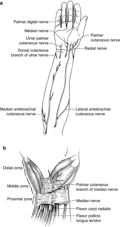
Figure 19.1.
(a) Cross section of the wrist. (d) The tunnel and its relations. From JE Skandalakis, GL Colborn, PN Skandalakis, et al. The carpal tunnel syndrome: Part I. Am Surg 58(1):72–76, 1992. Reprinted with permission from American Surgeon.
Figure 19.2.
The superficial and deep palmar arches and the topography of the motor branches of the median nerve. From JE Skandalakis, GL Colborn, PN Skandalakis, et al. The carpal tunnel syndrome: Part I. Am Surg 58(1):72–76, 1992. Reprinted with permission from American Surgeon.
Figure 19.3.
Superficial relations of the flexor retinaculum. (1) Radial artery, (2) flexor carpi radialis tendons, (3) palmaris longus tendon, (4) ulnar artery and nerve, (5) flexor carpi ulnaris tendon, (6) palmar cutaneous branch of median nerve, (7) palmar branch of radial artery, (8) three thenar muscles, (9) palmar cutaneous branch of ulnar nerve, and (10) three hypothenar muscles. Modified from JE Skandalakis, GL Colborn, PN Skandalakis, et al. The carpal tunnel syndrome: Part II. Am Surg 58(2):77–81, 1992 with permission from American Surgeon.
Figure 19.4.
The proximal surgical zone.
Figure 19.5.
(a) Palmar cutaneous branches of the ulnar, musculocutaneous, radial, and median nerves. (b) Palmar cutaneous branch of the median nerve. From JE Skandalakis, GL Colborn, PN Skandalakis, et al. the carpal tunnel syndrome: Part III. Am Surg 58(3):158–166, 1992. Reprinted with permission from American Surgeon.
Table 19.1
Upper proximal zone divisions
Ulnar side | |
Ulnar trio | Flexor carpi ulnaris |
Ulnar nerve | |
Ulnar artery | |
Central (median) area | |
Ulnar bursae with flexor digitorum superficialis and profundus tendons | |
Median duo
Stay updated, free articles. Join our Telegram channel
Full access? Get Clinical Tree
 Get Clinical Tree app for offline access

| |