and John E. Skandalakis1
(1)
Centers for Surgical Anatomy and Technique, Emory University School of Medicine Piedmont Hospital, Atlanta, GA, USA
Abstract
Few organs are more easily exposed and mobilized than are loops of small intestine; adhesions from previous surgery are the chief obstacles. Detailed step-by-step technique is given for jejunostomy and resection of small bowel for tumor. The surgeon should always be conservative. In benign disease, the ileocecal valve should be spared. There is no good way to identify an isolated loop of small intestine without following it in one direction to the duodenojejunal junction or in the other direction to the ileocecal junction.
An intussusception is created when a proximal segment of intestine (the intussusceptum) invaginates into the portion of intestine immediately distal to it (the intussuscipiens). Meckel’s diverticulum is the most common form of intussusception in children.
Anatomy
General Description of the Small Intestine
Information about the duodenum has already been presented in detail in Chap. 8. In this chapter, general descriptions of the intestine include the duodenum, but the primary focus here is on the jejunum and ileum.
Length of the Intestine
The length of the alimentary tract in humans has proven surprisingly difficult to measure. An average of 6–6.5 m for the small intestine has been widely quoted in textbooks. This figure bears an unknown relation to the length of the intestine in the living patient, since at death the intestines elongate; tonus is lost much faster in longitudinal muscle than in circular muscle. There is some evidence that intestinal length is greater in obese individuals.
The surgeon is more concerned with the length of intestine remaining after a resection than with the amount resected. Before the intestine is removed accurate measurements should be made with the least manipulation possible.
Dimensions of the Mesentery
The length of the mesentery measured between the attachment to the intestine and the root of the mesentery usually does not exceed 20–25 cm.
Layers of the Wall of the Intestine
The intestinal wall is composed of a serosa of visceral peritoneum, longitudinal and circular muscle, a submucosa of connective tissue, and a mucosa of connective tissue, smooth muscle, and epithelium (Fig. 10.1). The integrity of an anastomosis is greater if the submucosa is included.
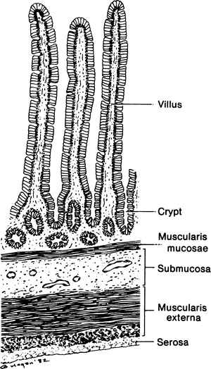
Figure 10.1.
Section through the wall of the small intestine. The submucosa should be included in stitches forming an anastomosis (By permission of JE Skandalakis, SW Gray, and JR Rowe Anatomical Complications in General Surgery. New York: McGraw-Hill, 1983).
Anatomy of the Ileocecal Valve
The ileocecal valve in most patients resembles the cervix protruding into the vagina or the pyloric opening into the duodenum (Fig. 10.2).
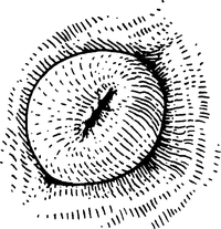
Figure 10.2.
The papillary appearance of the ileocecal valve in the living patient (By permission of JE Skandalakis, SW Gray, and JR Rowe. Anatomical Complications in General Surgery. New York: McGraw-Hill, 1983).
The closing mechanism of the papilla is formed by two rings of thickened circular muscle, one at the base of the papilla and one at the free end.
Vascular System of the Small Intestine
Arterial Supply
The intestinal vessels may be appreciated from Fig. 10.3. The superior mesenteric artery arises from the aorta below the origin of the celiac trunk.
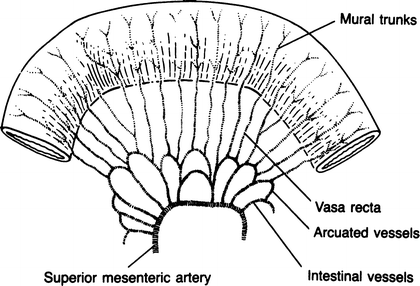
Figure 10.3.
Arterial supply to the small bowel (By permission of JE Skandalakis, SW Gray, AR Mansberger, et al. Hernia: Surgical Anatomy and Technique. New York: McGraw-Hill, 1989).
From the arches of the arcades, numerous arteries (the vasa recta) (Figs. 10.3 and 10.4) arise and pass (without cross-communication) to enter the intestinal wall. They may bifurcate to supply each side or they may pass singly to alternate sides of the intestine. Before piercing the muscularis externa, the vasa recta branch beneath the serosa, but do not anastomose. There is no collateral circulation between the vasa recta or their branches at the surface of the intestines. This configuration provides the best supply of oxygenated blood to the mesenteric side of the intestine and the poorest supply to the antimesenteric border.
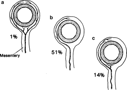
Figure 10.4.
(a) The vasa recta may divide into two short vessels to the mesenteric side of the intestine and two long vessels supplying the rest of the intestinal wall. (b) More frequently, a single, long vessel supplies one side of the intestine, alternating with a vessel supplying the other side. (c) A single long and a single short vessel serving one side only. The remaining 34 % are various combinations of paired, single, long, and short vessels (By permission of JE Skandalakis, SW Gray, and JR Rowe. Anatomical Complications in General Surgery. New York: McGraw-Hill, 1983).
Venous Drainage
The veins travel with the arteries in the mesentery to reach the superior mesenteric vein.
Stay updated, free articles. Join our Telegram channel

Full access? Get Clinical Tree


