and John E. Skandalakis1
(1)
Centers for Surgical Anatomy and Technique, Emory University School of Medicine Piedmont Hospital, Atlanta, GA, USA
Abstract
Surgery of the duodenum includes duodenotomy, duodenostomy, duodenal resection, pyloromyotomy, and partial anastomosis with stomach or jejunum. Good results from surgical procedures can be achieved if surgeons have good anatomical knowledge and if they practice good technique and conservative skeletonization. The duodenum is one of the most difficult areas to approach when operating, because the duodenum and pancreas are fixed; they share a common blood supply (superior and inferior pancreaticoduodenal arcades). The area of the opening of the common bile duct and pancreatic ducts is very complex.
Maneuvers for achieving exposure and mobilization of the duodenum are presented. A pancreaticoduodenectomy should be performed when malignant disease is found; with benign disease, a more conservative approach is the preferred treatment. The variations in the circular musculature must be kept in mind when performing pyloromyotomy. Step-by-step technique is given for repair of vascular compression of the duodenum.
Anatomy
General Description of the Duodenum
The first, or superior, portion of the duodenum (5 cm long) passes upward from the pylorus to the neck of the gallbladder. The proximal end is movable and enclosed by the same peritoneal layers that invest the stomach. The distal half of the first portion becomes retroperitoneal. Posteriorly, this portion is in intimate contact with the common bile duct, portal vein, and gastroduodenal artery. The duodenum is separated from the inferior vena cava by a small amount of connective tissue.
The second, or descending, portion (7.5 cm long) lies posterior to the transverse mesocolon and anterior to the right kidney and inferior vena cava. The left border is attached to the head of the pancreas. The common bile and pancreatic ducts open into the left side of this portion of the duodenum.
The third and fourth portions of the duodenum lie inferior to the transverse mesocolon. Their cranial surfaces are in contact with the uncinate process of the pancreas (Fig. 8.1). The parietal peritoneum covering the fourth portion of the duodenum contains folds beneath which are blind recesses or paraduodenal fossae.
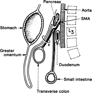
Figure 8.1.
Diagrammatic sagittal section showing the position of the duodenum in relation to the aorta and the superior mesenteric artery. The transverse mesocolon is marked by an X. SMA superior mesenteric artery (modified by permission of SW Gray, JT Akin, JH Milsap, et al. Contemp Surg 10:33–56, 1977).
The third, or horizontal, portion of the duodenum (10 cm long) passes to the left and slightly upward, crossing anterior to the inferior vena cava and posterior to the superior mesenteric artery and vein. The fourth, or ascending, portion (2.5 cm long) passes upward and slightly to the left, crossing the spine anterior to the aorta. It may cover the origin of the inferior mesenteric artery from the aorta. The duodenum ends at the duodenojejunal flexure, which usually lies immediately to the left of the aorta.
At the gastroduodenal junction, the continuity of the circular musculature is interrupted by a ring-shaped septum of connective tissue derived from the submucosa. Proximal to this ring, the circular muscle layer thickens to form the pyloric sphincter of the stomach; distal to the ring there is an abrupt decrease in the thickness of the circular muscle to form the relatively thin-walled duodenum. This decrease results in a pyloric “os pylorus” surrounded by a duodenal fornix. This arrangement must be kept in mind when performing pyloromyotomy.
The gastroduodenal junction is marked internally by the submucosal glands of Brunner; this may not correspond to the muscular junction. The submucosal glands may extend a few centimeters into the pylorus, and occasionally, antral gastric mucosa may prolapse through the pylorus to produce a radiological finding, but not a true clinical syndrome.
The duodenojejunal junction is marked externally by the attachment of the suspensory ligament of Treitz. This ligament or muscle is a fibromuscular band that arises from the right crus of the diaphragm, inserting on the upper surface of the duodenojejunal flexure. It passes posterior to the pancreas and the splenic vein and anterior to the left renal vein.
Usually the suspensory ligament inserts on the duodenal flexure and the third and fourth portions of the duodenum (Fig. 8.2b). Alternatively, it may insert on the flexure only (Fig. 8.2a), or on the third and fourth portions only (Fig. 8.2c), or there may be multiple attachments (Fig. 8.2d).
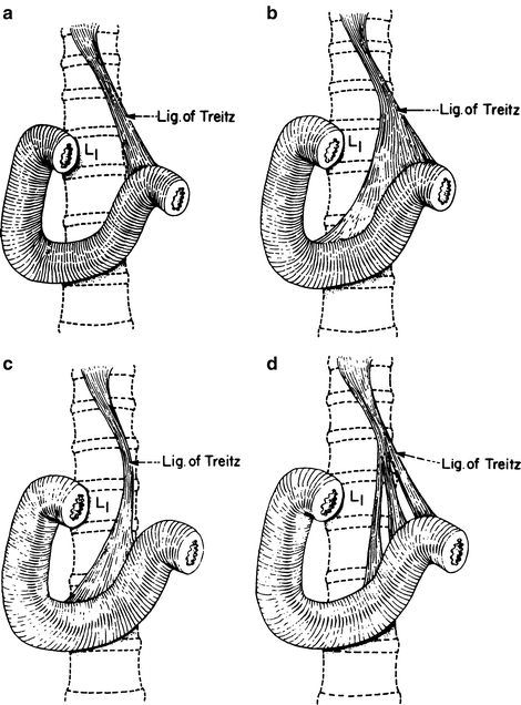
Figure 8.2.
Four configurations of the suspensory ligament of Treitz: (a) attachment to the duodenojejunal flexure. (b) Attachments to the flexure and the third and fourth portions of the duodenum. (c) Attachments to the third and fourth portions only. (d) Multiple separated attachments of the suspensory ligament (By permission of JE Skandalakis, JT Akin, JH Milsap, et al. Contemp Surg 10:33–56, 1977).
Vascular System of the Duodenum
Arteries
The blood supply of the duodenum is confusing due to the diverse possibilities of origin, distribution, and individual variations (Figs. 8.3, 8.4, and 8.5). This is especially true of the blood supply of the first portion of the duodenum.
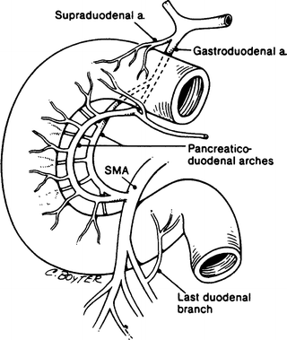
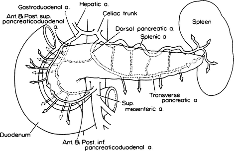
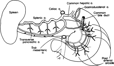
Figure 8.3.
Major arterial supply to the duodenum. SMA superior mesenteric artery. From SW Gray, GL Colborn, LB Pemberton, LJ Skandalakis, and JE Skandalakis. Am Surg 15(4):257–261; 15(5):291–298; 15(7):469–473; 15(8):492–494, 1989. Reprinted with permission from American Surgeon.
Figure 8.4.
Anterior view of arterial supply of the duodenum and pancreas (By permission of JE Skandalakis, SW Gray, JS Rowe, et al. Contemp Surg 15(5):17–40 and 15(6):21–50, 1979).
Figure 8.5.
Posterior view of arterial supply of the duodenum and pancreas (By permission of JE Skandalakis, SW Gray, JS Rowe, et al. Contemp Surg 15(5):17–40 and 15(6):21–50, 1979).
The first part of the duodenum is supplied by the supraduodenal artery and the posterior superior pancreaticoduodenal branch of the gastroduodenal artery (retroduodenal artery as described by Edwards, Michels, and Wilkie), which is a branch of the common hepatic artery.
Stay updated, free articles. Join our Telegram channel

Full access? Get Clinical Tree


