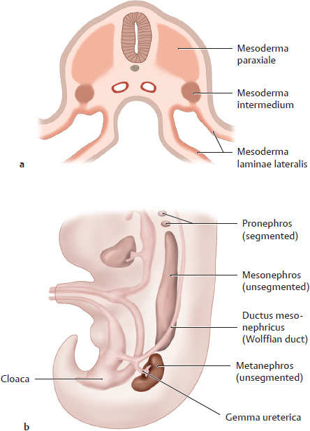
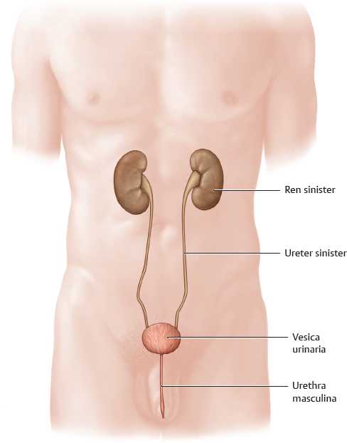
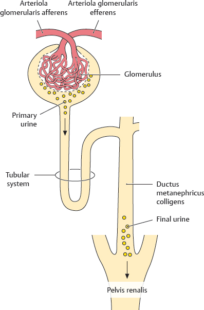
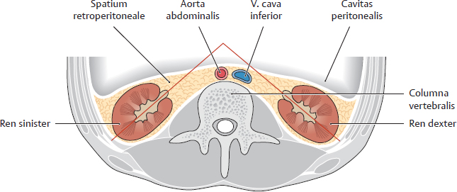
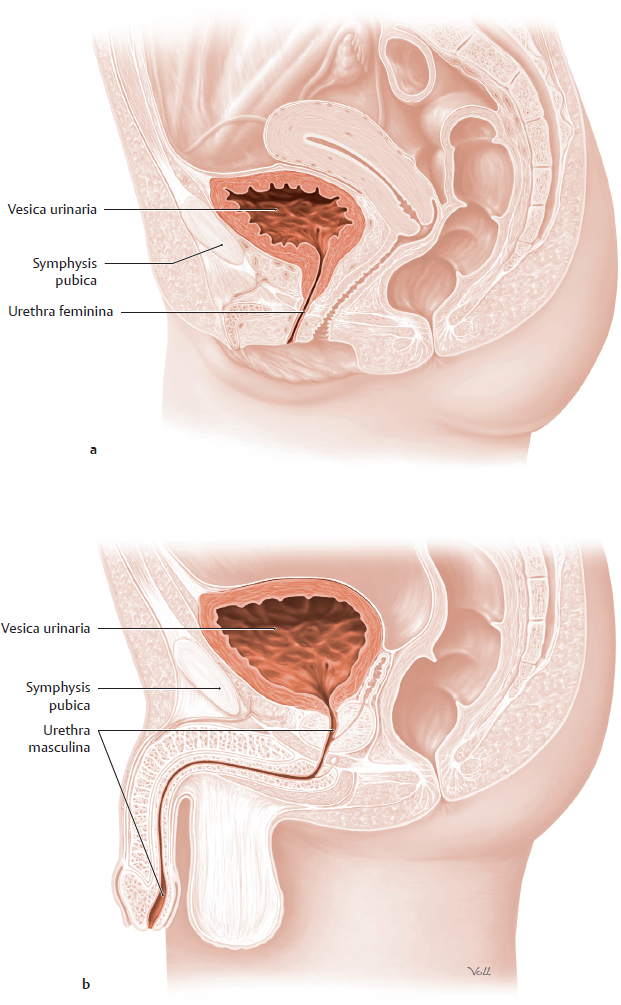
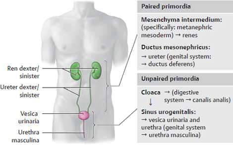
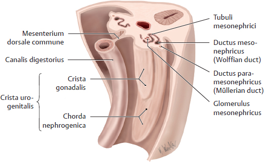
5. Urinary System Introduction The urinary organs extend from the abdomen through the pelvis. Since they are closely related to the genital organs, both groups of organs are often referred to collectively as urogenital organs. For didactic reasons, both systems will be discussed separately in the following chapters. The urinary organs help regulate the level of water and minerals in the body, and thus help regulate osmotic pressure. They excrete end products of body metabolism and harmful substances into a watery fluid, the urine (the excreted metabolites are dissolved in the water component of urine). By regulating the amount of water in the body, the renes also influence blood pressure. Through excreting or retaining sodium, potassium, calcium and chloride ions, they are involved in regulating the level of these important electrolytes in blood. In addition, the blood’s acid-base balance is influenced by the excretion or retention of hydrogen ions. Many pharmaceutical substances are excreted through the renes. The renes also influence blood pressure by producing the enzyme renin, and the formation of red blood cells by producing the hormone erythropoietin. Lastly, they also play an important role in vitamin D metabolism. A Overview of the urinary organs Male urinary organs, anterior view. The urinary system consists of the following organs: • the paired renes, which continuously produce urine; • the paired ureteres, which transport urine from the renes to the vesica urinaria; • the unpaired vesica urinaria, which temporarily stores and discharges urine in a controlled manner; and • the unpaired urethra. In women it is called the urethra feminina and it is solely a urinary organ, whereas in men it is called the urethra masculina and it is also a genital organ. In the urinary system, the urethra is involved in discharging urine from the vesica urinaria to the outside of the body. In men it is also serves as a passageway for sperm. B Basics of urine production The nephron, illustrated above, is the smallest functional unit of the renes (see p. 44). In the glomeruli, richly branched capillary loops, which are supplied by branches of the aa. renales, drain an ultrafiltrate of blood called primary urine into a system of tubules. Adults produce approximately 170 liters of primary urine in 24 hours. However, in the tubular system, the primary urine is concentrated to 1% of its volume (by reabsorption of electrolytes and water back into the blood), and based on its composition, further modified with electrolytes and hydrogen ions. The volume of final urine formed in 24 hours is 1–2 liters. The final urine drains through ductus metanephrici colligentes into the pelvis renalis and then it is carried by the ureteres to the vesica urinaria. C Location of the renes and ureteres Horizontal section of the body at the level of the first vertebra lumbalis, viewed from above. Both renes are embedded in a fatty, connective tissue capsule (capsula adiposa). They are located in the spatium retroperitoneale with one on either side of the columna vertebralis. The spatium retroperitoneale also contains the ureteres (not visible in this section), which extend downward to the pelvis minor to reach the vesica urinaria. The hilum of each ren faces medially and anteriorly (red axes). D Location of the vesica urinaria and urethra Midsagittal section of a female (a) and male (b) pelvis, each viewed from the left side. In both sexes, the vesica urinaria is located in the pelvis minor behind the symphysis pubica. In females it is situated in front of both the vagina and the uterus, and in males it is in front of the rectum. Depending on its degree of distension, the vesica urinaria is flattened or spherical. The vesica urinaria is straight and short, while the urethra masculina traverses the penis and bends multiple times along its course. A Overview of the embryonic development of the urinary organs The embryonic development of the urinary organs is complex and overlaps with the development of the genital and digestive organs: • overlap with the genital system: the development of some parts of the male reproductive system (see p. 52) is closely related to the development of the ductus mesonephrici, ureteres, and sinus urogenitalis. • overlap with the digestive system: the canalis analis is derived from the cloaca. The development of the urinary system can be divided into the development of the paired renes and ureteres, and development of the unpaired vesica urinaria and urethra. The renes and ureteres arise from the mesenchyma intermedium. The vesica urinaria and urethra develop from the sinus urogenitalis, which formed from the ventral portion of the cloaca in the region of the future pelvic floor (see p. 37). The sinus urogenitalis is derived from endoderma. Thus, the urinary organs are derived from two germ layers. Over the course of development the two sets of urinary organs will connect with each other. B The urogenital ridge (crista mesonephrica) Posterior body wall of the embryo, viewed from the front and above. The renal and internal genital primordia border each other. They bulge ventrally into the body cavity in the form of two ridges: the chorda nephrogenica and the crista gonadalis or crista mesonephrica. The developing gonadae lie anteromedially to the developing ren systems. The ductus paramesonephrici (Müllerian ducts), which in females develop into the tubae uterinae and uterus, lie anterolaterally to the renal primordia.
5.1 Overview of the Urinary System
5.2 Development of the Renes, Pelvis Renalis, and Ureteres
Basicmedical Key
Fastest Basicmedical Insight Engine