20. Organs of the Genital System and their Neurovasculature Classification of the genital organs The genital organs of the male and female can be classified in various ways: • Topographically (A) as – internal genital organs (internal genitalia) or – external genital organs (external genitalia) • Functionally (B, C) as – organs for germ-cell and hormone production (gonadae) or – organs of transport, incubation and copulation, plus accessory sex glands • Ontogenically (see p. 46) as – the undifferentiated gonad primordium (develops into the gonadae) – two undifferentiated duct systems (develop into the male and female transport organs, the female uterus, a portion of the female copulatory organ, and one of the accessory sex glands in the male) – the sinus urogenitalis and its derivatives (giving rise to the external genitalia of both sexes, the accessory sex glands, and portions of the copulatory organs) A Male and female internal and external genitalia*
20.1 Overview of the Genital Tract
| Male | Female |
Internal genitalia | Testis Epididymis Ductus deferens Prostata Gl. vesiculosa Gl. bulbourethralis | Ovarium Uterus Tuba uterina Vagina (upper portion) |
External genitalia | Penis and urethra Scrotum and coverings of the testis | Vagina (vestibulum vaginae only) Labia majora and minora Mons pubis Gll. vestibulares major and minor Clitoris |
* The female external genitalia (pudenda) are known clinically as the vulva. |
B Functions of the male genital organs
Organ | Function |
Testis | Germ-cell production Hormone production |
Epididymis | Reservoir for sperm (sperm maturation) |
Ductus deferens | Transport organ for sperm |
Urethra | Transport organ for sperm and urinary organ |
Accessory sex glands (prostata, gll. vesiculosae, and gll. bulbourethrales) | Production of secretions (semen) |
Penis | Copulatory and urinary organ |
C Functions of the female genital organs
Organ | Function |
Ovarium | Germ-cell production Hormone production |
Tuba uterina | Site of conception and transport organ for zygote |
Uterus | Organ of incubation and parturition |
Vagina | Organ of copulation and parturition |
Labia majora and minora | Copulatory organ |
Gll. vestibulares major and minor | Production of secretions |
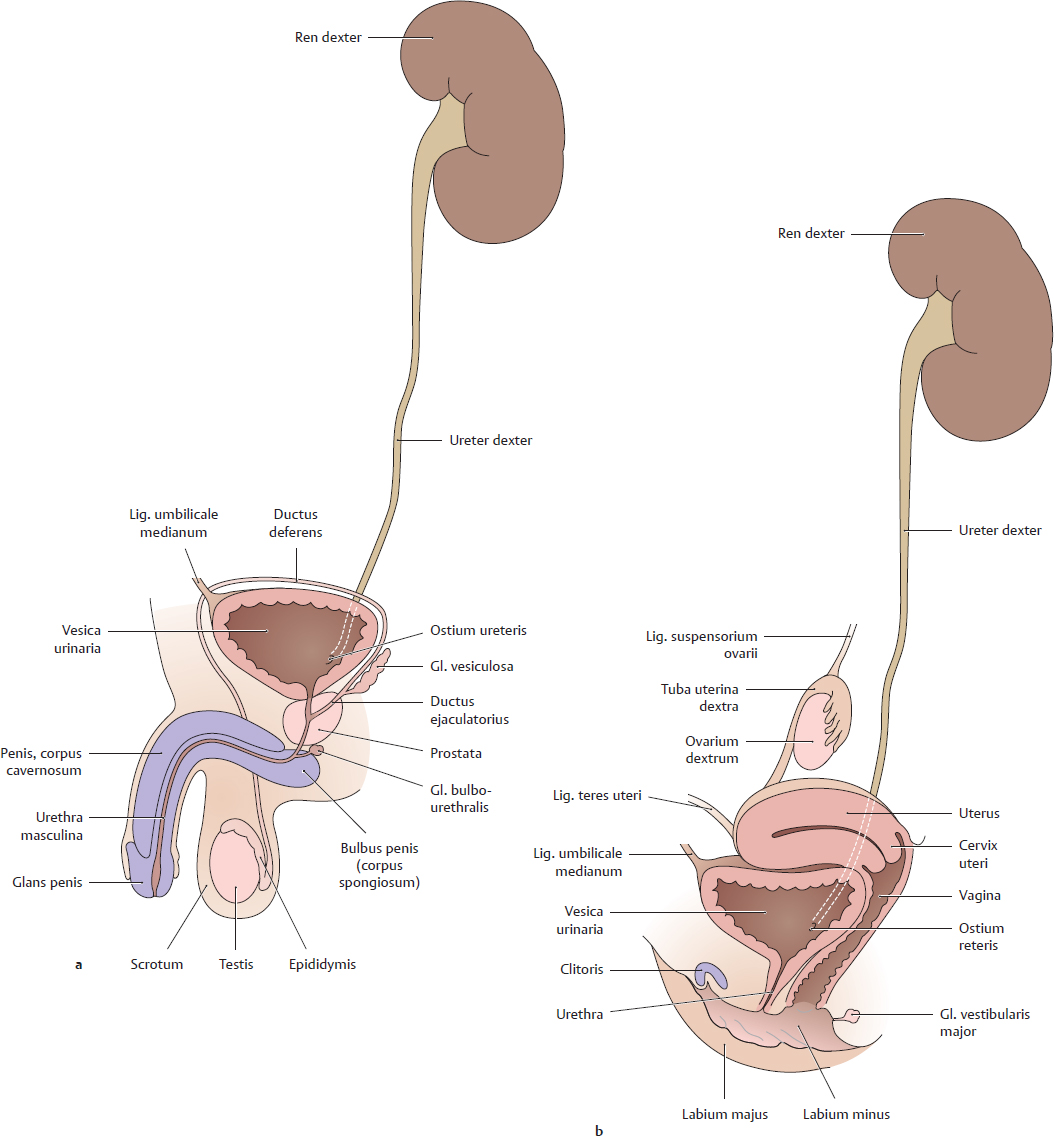
D Overview of the urogenital system
Schematic representation of the urogenital apparatus in the male and female, viewed from the left side. Unpaired pelvic organs and the external genitalia are shown in midsagittal section.
a In the male, the urinary and genital organs are closely interrelated functionally and topographically. The urethra passes through the prostata, which is derived embryologically from the urethral epithelium. All of the accessory sex glands (prostata, gll. vesiculosae, and gll. bulbourethrales) ultimately discharge their secretions into the urethra.
b In the female, the urinary and genital tracts are functionally separate from each other. Topographically, however, the anterior wall of the uterus is closely related to the vesicula urinaria. In the external genital region as well, the urethra is embedded in the paries anterior of the vagina.
For these reasons, the collective term urogenital system is generally used.
20.2 Female Internal Genitalia: Overview
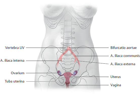
A Projection of the female internal genitalia onto the pelvis
Anterior view. The bifurcatio aortae into the aa. iliacae communes is also shown to aid orientation. The uterus, like the vagina, is located in the pelvic midline while the ovaria are superior, lateral, and posterior to the uterus in the RLQ and LLQ. Each ovarium occupies a fossa located just inferior to the division of the a. iliaca communis. The tubae uterinae do not pass to the ovaria by the shortest route but circle around them from the lateral side, because both of the ductus paramesonephrici (which develop into the tubae uterinae) run lateral to the crista gonadalis in which the ovaria develop.

B Uterus and vagina: Relationship to the pelvic organs
Midsagittal section through a female pelvis, viewed from the left side. The peritoneum has been outlined in color. The uterus directly overlies the vesica uterina, and the rectum is posterior to the uterus. The fundus and corpus (body) of the uterus are covered by peritoneum viscerale, which is reflected onto the vesica urinaria and rectum to form the excavatio vesicouterina and excavatio rectouterina. The peritoneum extends farther down the posterior wall of the uterus than its anterior wall, with the result that the posterior part of the cervix uteri and upper vagina is covered by peritoneum while the anterior part is not. The vagina is surrounded on all sides by pelvic connective tissue. This tissue is thickened anteriorly and posteriorly to form the septa vesicovaginale and rectovaginale.
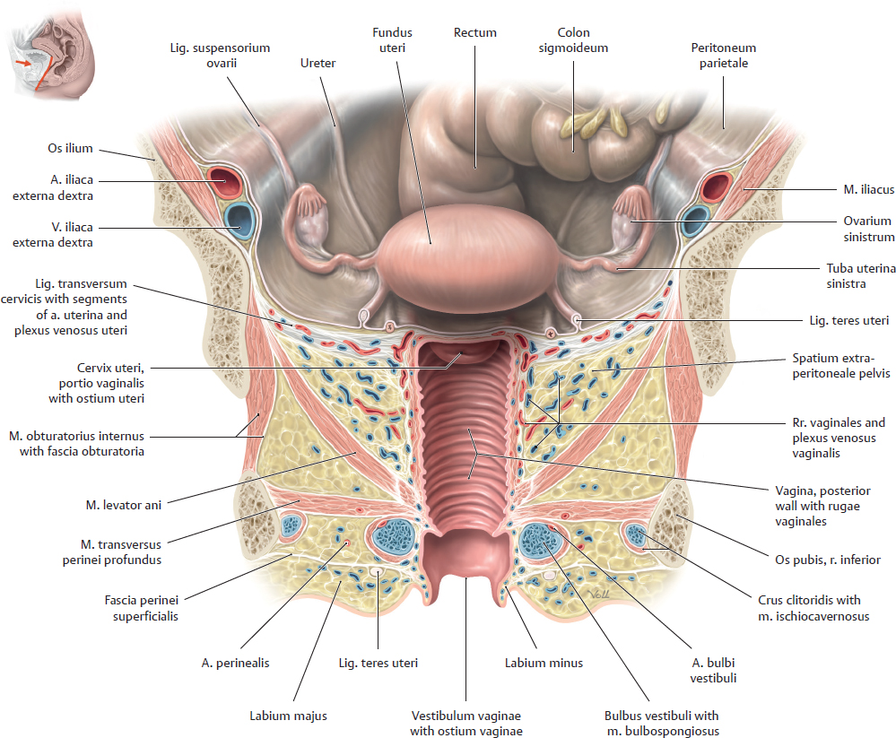
C The female genital organs in situ
Slightly angled coronal section, anterior view. The vesica urinaria, which lies anterior to the vagina and inferior to the fundus uteri (see B), is not shown. This illustration represents a compilation of multiple sections to provide a single integrated view. The fundus uteri, which is directed anteriorly owing to its anteverted and anteflexed position (see p. 318), projects out of the deeper plane of section toward the observer. Around the vagina is a connective-tissue space containing an elaborate venous plexus. This loose connective tissue allows for considerable expansion of the vagina during childbirth. The sections of arterial vessels are arterial rr. vaginales as well as sections of the aa. vesicales inferiores.
20.3 Female Internal Genitalia: Topographical Anatomy and Peritoneal Relationships; Shape and Structure
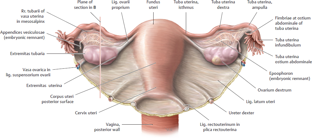
A Uterus and adnexa: Topography and peritoneal relationships
Posterosuperior view of the uterus, adnexa, and the posterior surface of the lig. latum uteri. The uterine adnexa (ovarium and tuba uterina) are attached to the superior border and posterior surface of the lig. latum uteri by folds of peritoneum (mesovarium and mesosalpinx, see B). The mesometrium, which follows the anteflexed position of the uterus, attaches the uterus to the pelvic sidewall and transmits the uterine vascular structures. The ovarium receives its vascular supply through the lig. suspensorium ovarii (these and other ligaments are reviewed in C). Note: The ureteres descend in the retroperitoneum to the base of the lig. latum uteri and run forward between its layers to the vesica urinaria, passing inferior to the a. uterina (not visible here, see p. 343). This relationship must be duly noted in operations on the uterus and lig. latum uteri (risk of ureteral injury).
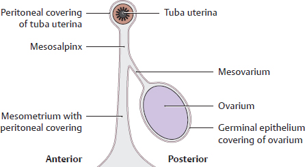
B Folds of peritoneum on the female genital organs (after Graumann, von Keyserlingk, and Sasse)
Sagittal section through the lig. latum uteri. The ovarium, tuba uterina, and much of the uterus (see A) are covered by peritoneum. The tuba uterina is attached to the superior margin of the lig. latum uteri by the mesosalpinx. The ovarium is attached to the posterosuperior surface of the lig. latum uteri by its own peritoneal structure, the mesovarium. These peritoneum-covered bands of connective tissue perform the same functions for the genital organs as the mesenteria do for the bowel and are named accordingly (see C): the mesovarium for the ovarium, the mesosalpinx for the tuba uterina (salpinx), and the mesometrium for the uterus. Collectively they form the lig. latum uteri.
C Ligaments and peritoneal structures of the female genital organs
Ligamentum latum uteri | Broad fold of peritoneum extending from the lateral pelvic wall to the uterus (transmits vascular structures to the internal genital organs). The ligament has three main parts that extend to specific organs: • Mesometrium = to the uterus • Mesosalpinx = to the tuba uterina • Mesovarium = to the ovarium The connective-tissue space between the two peritoneal layers of the lig. latum uteri is known clinically as the parametrium |
Ligamentum transversum cervicis (ligamentum cardinale) | Transverse bands of connective tissue between the cervix uterina and pelvic wall (paracervix) |
Ligamentum teres uteri | Distal remnant of the gubernaculum (embryonic cord in both sexes, guides the descent of the testis or ovarium). Extends from the lateral angle of the uterus through the canalis inguinalis into the subcutaneous connective tissue of the labium majus |
Plica rectouterina | Peritoneum-covered fold of connective tissue between the uterus and rectum; often contains smooth muscle (m. rectouterinus) |
Ligamentum ovarii proprium | Proximal remnant of gubernaculum passing from the extremitas uterina of the ovarium to the angle of the uterus with the tuba uterina |
Ligamentum suspensorium ovarii | Fold of peritoneum stretching from the pelvic wall to the ovarium; transmits the ovarian vessels |
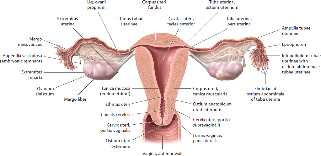
D Uterus and tubae uterinae: Shape and structure
Posterior view of a coronal section with the uterus straightened and the mesometrium removed. The uterus consists basically of the corpus (with the fundus) and cervix, the corpus being joined to the cervix by a narrow isthmus approximately 1 cm long. Macroscopically, the isthmus uteri is classified as part of the cervix but histologically it is lined by endometrium. The junction of the corpus and cervix uteri is located at the ostium anatomicum uteri internum of the uterus. The lumen of the uterus, called the cavitas uteri, communicates with the vaginal lumen through the isthmus uteri and canalis cervicis uteri. It has a total length (“probe length”) of 7–8 cm. The cavitas uteri presents a triangular shape in coronal section. The cervix uteri is subdivided into a portio supravaginalis cervicis and portio vaginalis cervicis. The ostium uteri is the opening in the portio vaginalis cervicis that is directed toward the vagina. The portio vaginalis cervicis projects into the vagina, forming recesses called the fornices vaginae.
The tuba uterina (total length approximately 10–18 cm) is subdivided from lateral to medial into the infundibulum, ampulla, and isthmus tubae uterinae, and pars uterina tubae uterinae. The ostium abdominale tubae uterinae at the infundibulum is surrounded by fimbriae (the “fimbriated end”) and opens into the cavitas peritonealis. The ostium uterinum tubae uterinae opens into the cavitas uteri.
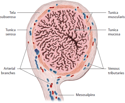
E Tuba uterina in cross-section: Wall structure
Cross-sectional view of the ampulla of a right tuba uterina. The mesosalpinx extends inferiorly. The three wall layers are clearly distinguishable (wall thickness = 0.4–1.5 cm):
• The tunica mucosa is raised into a great many folds that occupy most of the tubal lumen. These folds are of key importance in transporting the zygote to the uterus. Postinflammatory adhesions between the mucosal folds may hamper or even prevent transport of the fertilized ovum (see p. 326).
• The tunica muscularis consists of several thin layers of smooth muscle that provide the tuba uterina with its motility (see B, p. 324) and propel the zygote toward the uterus by a ciliated epithelium.
• The tunica serosa (peritoneal covering) of the tuba uterina is continuous with the mesosalpinx.
20.4 Female Internal Genitalia: Wall Structure and Function of the Uterus
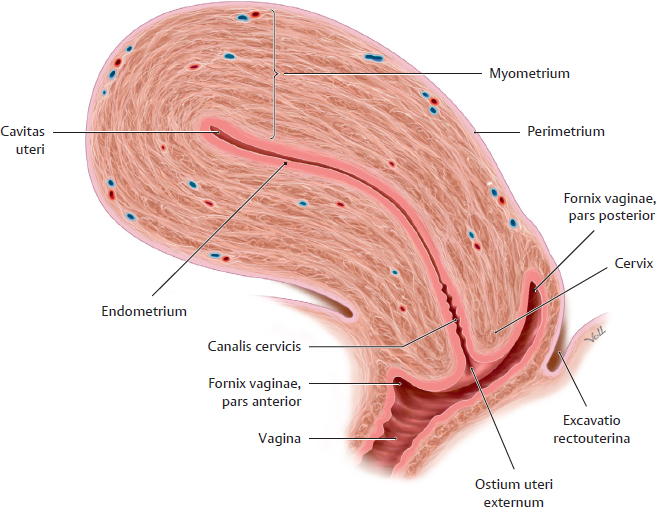
A Longitudinal section through the uterus
Viewed from the left side.
B Wall structure of the uterus
The uterine wall also consists of three layers from inside to outside:
• Tunica mucosa or endometrium (see D): Single layer of columnar epithelium (epithelial layer) on a connective-tissue base (lamina propria)
• Tunica muscularis or myometrium (see C):
Several smooth-muscle layers with a total thickness of approximately 1.5 cm
• Tunica serosa or perimetrium: Tunica serosa covering the anterior and posterior sides of the corpus uteri and the posterior wall of the cervix uteri. The tela subserosa adjacent to the myometrium becomes adventitia in areas where the uterus lacks a peritoneal covering (e.g., at the attachment of the lig. latum uteri).
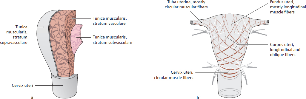
C Layers (a) and functional principle (b) of the myometrium (after Rauber and Kopsch)
The myometrium (tunica muscularis) of the uterus consists of three layers from outside to inside:
• Stratum supravasculare: thin outermost layer with criss-crossing lamellae; stabilizes the uterine wall
• Stratum vasculare: thick intermediate layer with a reticular pattern of muscle fibers; very vascular; the principal source for uterine contractions during labor
• Stratum subvasculare: thin innermost layer just below the endometrium; provides for functional closure of the ostium uterinum tubae uterinae. Its contraction promotes separation of the uterine tunica mucosa (shedding of the functional layer) during menses and separation of the placenta after childbirth.
The myometrium performs two seemingly contradictory functions: It must keep the uterus closed during pregnancy, but it must open the cervix during childbirth. To fulfill these functions, the individual muscle layers (see above) are equipped with longitudinal, oblique, and transverse or circular fibers. The circular muscle fibers are most abundant in the cervical region and serve to maintain closure of the cervix during pregnancy. The longitudinal and oblique muscle fibers are most abundant in the corpus and fundus uteri; they shorten the uterus and lower the fundus during childbirth. The myometrium blends with the circular fibers of the uterine tube muscles at the fundus uteri near the ostium uterinum tubae uterinae. Myometrial contractions are stimulated most effectively by the pituitary hormone oxytocin. These contractions occur not only during labor and delivery but also during menstruation, when they aid in expulsion of the uterine mucosa. Benign tumors of the myometrium (fibroids, myomas) may cause abnormalities of menstrual bleeding.
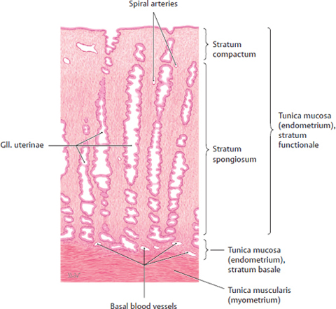
D Structure of the uterine mucosa (endometrium)
Structurally, the endometrium consists of a simple columnar epithelial cell layer and a lamina propria. The epithelial layer lines the uterine surface and encloses the tubular, coiled endometrial gll. uterinae. The lamina propria, which surrounds and supports the gll. uterinae, is made up of connective tissue (stroma) and the vessels embedded in it. The endometrium is functionally subdivided into a basal layer (stratum basale) and a functional layer (stratum functionale). The stratum basale is approximately 1 mm thick, is largely exempt from the cyclical changes in the endometrium, and is not shed during menstruation. The stratum functionale varies in thickness at different phases of the ovarian cycle in women of reproductive age. It is shed at intervals of approximately 28 days during menstruation. It is thickest during the secretory phase of the ovarian cycle, at which time it consists of a superficial stratum compactum and a deeper stratum spongiosum. It receives its blood supply from tortuous vessels called spiral arteries. While in this secretory state, the endometrium is most receptive to the implantation of a zygote. The tunica mucosa of the cervix uteri does not participate in these cyclical changes.
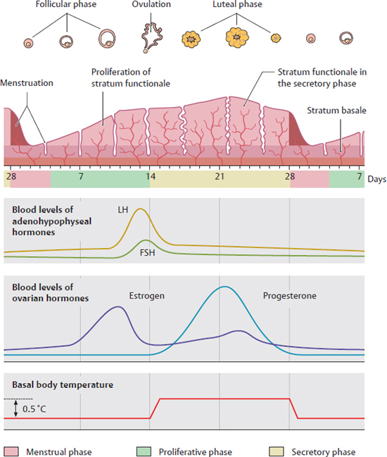
E Cyclical changes in the endometrium
The ovarium secretes estrogens (e.g., estradiol) and progestins (e.g., progesterone) on a cyclical basis. Estrogens stimulate proliferation of the endometrium, while progestins induce its secretory transformation. The release of both hormones is controlled chiefly by the hormones FSH (follicle stimulating hormone) and LH (luteinizing hormone), which are secreted cyclically by the hypophysis. While estrogens are produced by the folliculus ovaricus, progestins are produced in significant amounts only by the corpus luteum. If conception does not take place, the corpus luteum regresses and stops producing hormones. As a result of this, the stratum functionale of the endometrium breaks down and is expelled during menstruation. Estrogen production by a new, hypophysis-stimulated folliculus ovaricus initiates a new cycle, which lasts an average of 28 days (1 lunar month). Ovulation usually occurs on day 14 of the cycle.
Note: For practical reasons, the first day of the menstrual period (which lasts about 4 days) is considered day 1 of the cycle, despite the fact that the cycle ends with menstruation. This is because the sudden onset of menstrual bleeding is easier to detect than its more gradual cessation. From the standpoint of the endometrium, however, the last day of the menstrual period (difficult to detect) marks the end of the cycle.
20.5 Female Internal Genitalia: Positions of the Uterus and Vagina
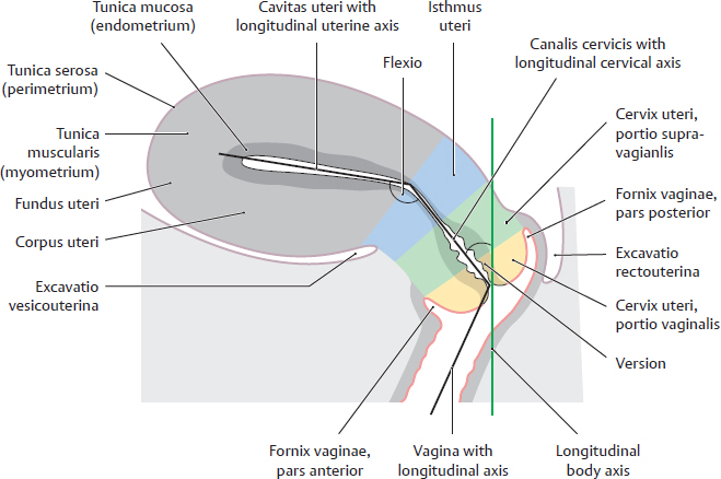
A Curvature and position of the uterus
Midsagittal section of the uterus and upper vagina, viewed from the left side.
Note the two angles that determine the normal anteversion and anteflexion of the uterus (see D). Posterior angulation and curvature of the uterus (retroflexion, retroversion) are considered abnormal. A retroverted uterus is more susceptible to descent because it is more closely aligned with the longitudinal axis of the vagina. Moreover, a retroverted uterus that enlarges during pregnancy may become immobile below the promontorium ossis sacri (L 5/S1 junction) and jeopardize the further course of the pregnancy by constraining uterine expansion.
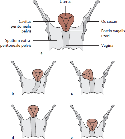
B Position and level of the uterus in the pelvis
Coronal section of the pelvis, anterior view. The uterus has been slightly straightened for clarity. Normally the uterus is located approximately in the median plane (a) with its portio vaginalis level with a line connecting the two spinae ischiadicae. The uterus may be displaced from this position to the left or right (sinistroposition or dextroposition, b and c) or may lie above or below the plane of the spinae ischiadicae (elevation or descent, see d and e). Anterior and posterior displacement (anteposition, retroposition) may also occur but are not illustrated here. Descent of the uterus usually results from a structural weakness of the diaphragma pelvis (chiefly the m. levator ani, often after numerous vaginal deliveries). Displacement of the uterus may cause complaints and functional disturbances due to pressure on adjacent organs (vesica urinaria, rectum). Descent of the uterus may even cause the portio vaginalis of the uterus to protrude from the vagina (cervical prolapse).
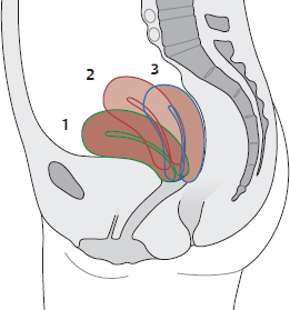
C Physiological changes in uterine position
Midsagittal section of the pelvis, viewed from the left side. Uterine position is directly affected by the varying degrees of vesica urinaria and rectal distention. 1 Vesica and rectum empty; 2 vesica and rectum full; 3 vesica full, rectum empty.
D Describing the position of the uterus in the pelvis
The position of the uterus in the pelvis can be described in terms of version, flexion, and position (angles are shown in A).
Version | Inclination of the cervix in the cavitas pelvis; defined by the angle between the cervical axis and the longitudinal axis of the body; the normal condition is anteversion |
Flexion | Inclination of the corpus uteri relative to the cervix; defined by the angle between the longitudinal axes of the cervix and corpus uteri; the normal condition is anteflexion |
Position | Position of the portio vaginalis of the uterus in the cavitas pelvis; physiologically, the portio vaginalis of the uterus is at the level of the interspinous line at the center of the pelvis |
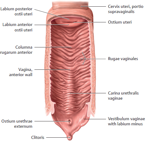
E Vagina
Posterior view. The vagina has been cut open along a coronal plane angled slightly posteriorly to display its paries anterior. The vaginal lumen presents an H-shaped cross section (see F), but the lumen in this dissection has been stretched open to a more circular shape (in situ the parietes posterior and anterior are closely apposed). The vaginal tunica mucosa has numerous transverse folds (rugae vaginales) as well as columnae rugarum anterior and posterior formed by the extensive venous plexus in the vaginal wall. The closely adjacent urethra raises the lower paries anterior of the vagina into a prominent longitudinal ridge (carina urethralis vaginae).
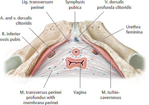
F Location of the vagina in the pelvic floor
This drawing illustrates the close proximity of the vagina and urethra. Muscular fibers from the m. transversus perinei profundus encircle the vagina.
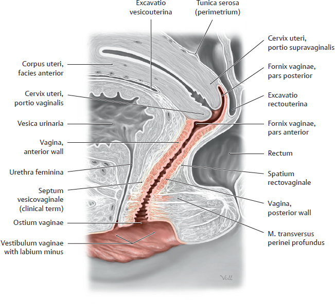
G Location of the vagina in the pelvis
Midsagittal section through a female pelvis, viewed from the left side. The longitudinal axis of the vagina is directed posterosuperiorly. The vagina is attached to the pelvic connective tissue anteriorly (septum vesicovaginale), posteriorly (septum rectovaginale), and laterally (not shown here). The fornix vaginae surrounds the portio vaginalis of the cervix, which itself is directed superiorly and anteriorly. As a result, the pars anterior fornicis vaginae is considerably lower than the pars posterior. The peritoneum viscerale extends far down the posterior uterine wall, bringing the pars posterior of the fornix vaginae into close proximity to the excavatio rectouterina (cul-de-sac, the lowest part of the female cavitas peritonealis).
20.6 Female Internal Genitalia: Epithelial Regions of the Uterus
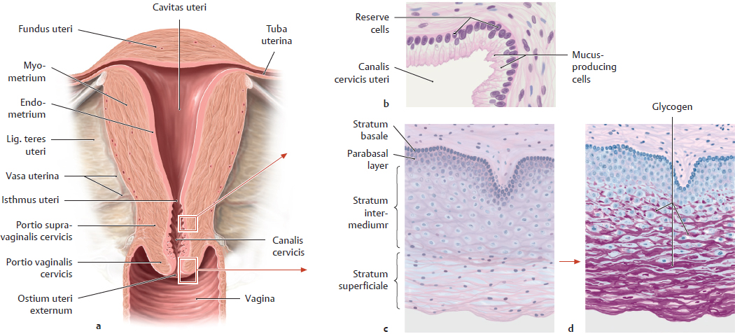
A Epithelial regions of the uterus
a Coronal section through uterus, anterior view; b–d Higher magnification of a: b Mucus-secreting columnar epithelium of the canalis cervicis uteri; c Squamous epithelium of the portio vaginalis cervicis; d Periodic acid–Schiff (PAS) method used to display glycogen (after Luellmann).
The cervix is the lower part uteri. It commences distal to the isthmus of the uterus as the portio supravaginalis cervicis, which is the upper part of the cervix surrounded by connective tissue of the parametrium, and ends at its lower portion that extends into the vagina (portio vaginalis cervicis). At the level of the portio supravaginalis cervicis the uterus is held in place by ligaments (mainly the lig. cardinalis, cf. p. 388). The tubular lumen of the cervix is called the canalis cervicis uteri. It is lined with tunica mucosa and begins at the ostium uteri internum and ends with the ostium uteri at the portio vaginalis of the cervix. The mucosal epithelium of the canalis cervicis uteri is composed of a single layer of mucus-secreting columnar epithelium. A parallel arrangement of folds (plicae palmatae), which form crypts, gives the epithelium a rough appearance on the surface. The function of the reserve cells located at the base of the epithelium is to replenish the cervical epithelium. Unlike the one-layered cervical epithelium, the vagina is lined with stratified, non-keratinized squamous epithelium, which depending on the hormonal situation in women may continue to the surface of the portio vaginalis of the cervix. The boundaries of the two epithelial layers can be either endo- or ectocervical (see C).
The stratified, nonkeratinized squamous epithelium lining the vagina (and portio vaginalis cervicis) is composed of up to 20 layers of cells and is made up of four tiers: strata basale, parabasale, intermedium, and superficiale. Typically, cells of the two superficial-most layers contain abundant glycogen as a result of differentiation. The epithelium exhibits cyclical changes: whereas at the preovulatory stage, all layers are well developed, at the postovulatory stage cells of the strata superficiale and intermedium desquamate and disintegrate. Glycogen that is released as a result nourishes lactic acid bacteria (lactobacillus acidophilus, Döderlein’s bacillus), which inhabit the vagina. The transformation of glycogen into lactic acid leads to the acidic vaginal milieu (pH 4–5), which mainly in the second half of the menstrual cycle protects against pathogens (see B). The slightly alkaline cervical mucus has a similar effect as a physiologic barrier against infection. For much of the menstrual cycle, it has a stretchy texture and seals the canalis cervicis uteri with a protective plug (barrier against ascending germs). The mucus is thin only at the time of ovulation and thus becomes penetrable by sperm.
B Defense mechanisms of the vagina and potential dysfunctions
The cavitas peritonealis has an anatomical communication with the exterior of the body (via vagina–canalis cervicis uteri–cavitas uteri– tuba uterina). This exposes the female to ascending infections, and this is why physiologic barriers against infection exist in the form of vaginal defense mechanisms. Dysfunction of these mechanisms may lead to gynecological inflammation and an increased risk of miscarriage.
Protection mechanism | Dysfunction due to |
• Physiological acidic vaginal milieu with a pH 4–5 • Effect of estrogens: stimulates vaginal epithelium proliferation and differentiation (glycogen storage) • Effect of progestins: leads to desquamation of superficial and intermediate vaginal cells • Conversion of glycogen into lactic acid through lactobacillus acidophilus (Döderlein’s bacillus) | • Elevated pH level: alkalizing effect of menstrual blood/cervical mucus • Lack of glycogen: lack of endogenous estrogen/progestins (childhood/old age/diseases) • Drugs: antibiotics disrupt vagina’s normal flora • Exogenous effects: sex life, tampons, improper anal hygiene, using alkaline soaps • Infections: colpitis especially caused by chlamydia, trichomonads and fungi (candida albicans) |
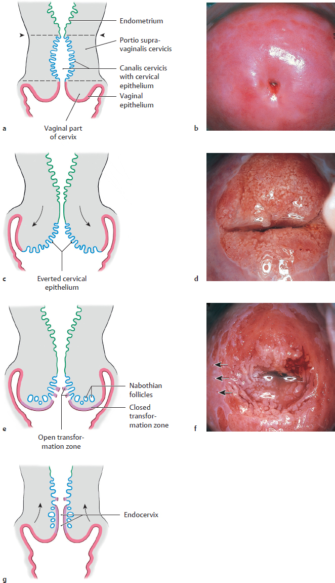
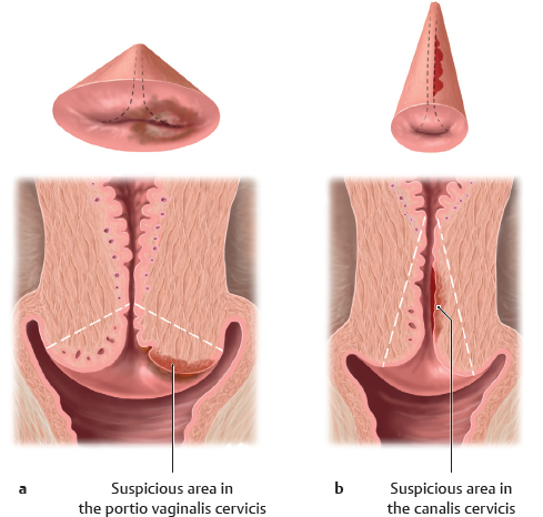
C Cervix uteri: Epithelial regions in pre-reproductive and post-reproductive stages
a, c, e, and g: Highly schematic coronal sections of the uterus and vagina, anterior view; colposcopic images of the portio vaginalis of the cervix before the onset of puberty (b nullipara) and during the reproductive phase (d and f multipara); b, d, and f from Nauth, H.F.: Gynäkologische Zytodiagnostik. Thieme, Stuttgart 2002).
The arrowheads mark the location of the ostium uteri internus; the dashed lines mark the boundaries of the canalis cervicis uteri (a). The boundary between the unilayered mucus-secreting columnar epithelium (cervical epithelium) of the canalis cervicis uteri and the stratified nonkeratinized squamous epithelium of the portio vaginalis cervicis and the vagina varies according to the woman’s hormonal status (see below). The visible portion of the uterus is called the ectocervix and the non-visible portion is called the endocervix.
Before the onset of puberty (a and b): Before the onset of the reproductive phase, the portio vaginalis cervicis is covered with squamous epithelium, the ecto-endocervical boundary is located within the canalis cervicis uteri (above the ostium uteri), thus it is not visible from the vagina.
During the reproductive phase (c–f): In response to hormonal stimuli (estrogen) the cervical tunica mucosa is everted and moves inside the vagina. It appears as a glandular field with a very rough surface on the ectocervix (d). The sharp boundary with the smooth pink-colored squamous epithelium of the portio vaginalis cervicis is thus located outside of the ostium uteri and is clearly visible from the vagina. The eversion of the cervical glandular field is believed to be related to higher fertility (easier for spermatozoa to enter the cervix). The columnar endocervical epithelium, which has everted onto the ectocervix adjusts to the altered vaginal milieu (acidic milieu in contrast to alkaline milieu of the canalis cervicis uteri) by converting to stratified nonkeratinized squamous epithelium (metaplasia). In this way, it is similar in structure and cyclic behavior to the regular squamous epithelium of the portio vaginalis cervicis. As the mucus-secreting cervical epithelium transforms into squamous epithelium the columnar glands become sealed over (closed transformation zone in contrast to open transformation zone where the glands are not overgrown and occluded, arrows in f point to the “open” orifices) leading to the formation of macroscopically visible mucus-filled retention cysts (Nabothian cysts), which are not considered problematic. Squamous epithelium in the transformation zone may become malignant and contribute in pre-stages (precancerous) to squamous cell carcinoma formation (see p. 322 f).
During postmenopause (g): Lower estrogen levels toward the end of the reproductive phase cause the relocation of cervical epithelium and the boundary of the endo- and ectocervix moves back into the canalis cervicis uteri (similar clinical presentation to b although the cervix changes shape after vaginal delivery).
20.7 Female Internal Genitalia: Cytologic Smear, Conization; Cervical Carcinoma
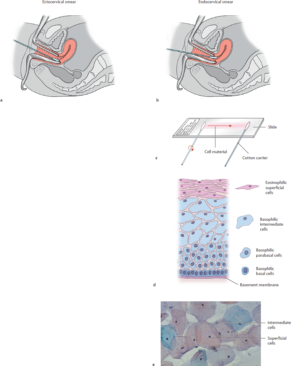
A Cytologic smear: Cell morphology of vaginal and vaginal part of cervical epithelium; early detection of cervical cancer
a and b Cytologic smears taken from the portio vaginalis cervicis and the canalis cervicis uteri, c transfer of smear material onto glass slide; d histological structure and cell morphology of the vaginal and cervical epithelium in the cytologic smear; e Pap staining of superficial and intermediate cells (e from Nauth H.F.: Gynäkologische Zytodiagnostik. Thieme, Stuttgart 2002).
Especially in the transformation zone of the cervix uteri, where with the onset of puberty the unilayered columnar epithelium of the cervix changes into stratified squamous epithelium (see p. 321), the squamous epithelium may undergo malignant transformation and turn into invasive squamous cell carcinoma. Because cervical cancer usually develops slowly over time, it is possible to detect it in its early stages with the help of cytologic smears. Thus, cytodiagnosis is one of the most important tools in early detection of cervical cancer (see D). Cytology testing is an obligatory part of the initial gynecologic examination and cancer screening (in Germany starting at the age of 20) and is also performed to evaluate suspicious changes in cervical tissue. The cytologic smear should always include cells from the uppermost epithelial layer, which if they are normal show signs of differentiation (see p. 320).
Two smears are taken routinely: the first one (a) must be taken at the surface of the portio vaginalis cervicis (ectocervix), and the second one (b) from the canalis cervicis (endocervix). The smear material is taken with a cotton swab and transferred to a slide and fixed (c). After that, the Papanicolaou method (known as Pap stain) is used to stain the cervical smear and evaluate it for characteristics of cell differentiation (cell shape, nuclear shape, nucleus-plasma ratio, see D). Because structure, height and the degree of maturation vary with the hormonal status (menstrual cycle) of a woman, it is crucial to confirm at which point during the cycle the smear is taken. If it is taken during the follicular phase (effect of estrogen), it is dominated by eosinophilic superficial cells, which are stained red, and flat, basophilic intermediate cells with pyknotic nuclei, which are stained greenish-blue (e). This proliferative phase, during which the uppermost cell layer is constantly regenerated by cells from the basal layer, normally lasts about a week. The postovulatory phase is dominated by cell differentiation and desquamation. Hence, the epithelium is thinner in the second part of the menstrual cycle.
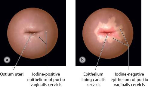
B Schiller’s iodine test for localization of abnormal epithelial regions
a Normal vaginal cervical epithelium stained by iodine produces a dark brown color; b Iodine negative, insufficiently differentiated cervical epithelium.
After inserting a speculum into the vagina, the portio vaginalis cervicis is initially examined macroscopically, if necessary with the help of a colposcope allowing a 6–40 fold magnification of the cervix. For the localization of abnormal areas, Schiller’s iodine test utilizes the glycogen content of the surrounding normal squamous epithelium. To that end, iodine solution (Schiller’s iodine test) is applied to the surface of the cervix. Normal squamous epithelium, regardless of whether is autochthonous or metaplastic, takes on a dark brown color. However, insufficiently differentiated squamous epithelium with a high or low glycogen content turns only light brown or is iodine negative. Thus, the iodine-unstained areas correspond to the location of undifferentiated epithelium. Iodine-negative areas are not specific but combined with suspicious cytology results (see above cytologic smear) of the same area, point to epithelial abnormalities. Hence, Schiller’s iodine test provides a method with which to assess the localization and expansion of cervical changes. Conization (see C) is used to remove affected areas.