
Diagnostic Checklist
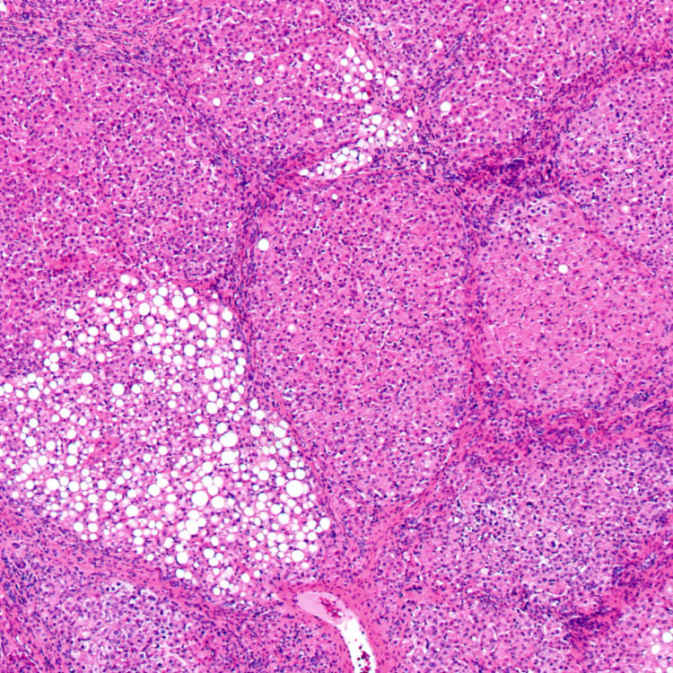
This liver with micronodular cirrhosis demonstrates focal areas of large droplet fatty change in some nodules. The fatty change in tyrosinemia is often patchy.
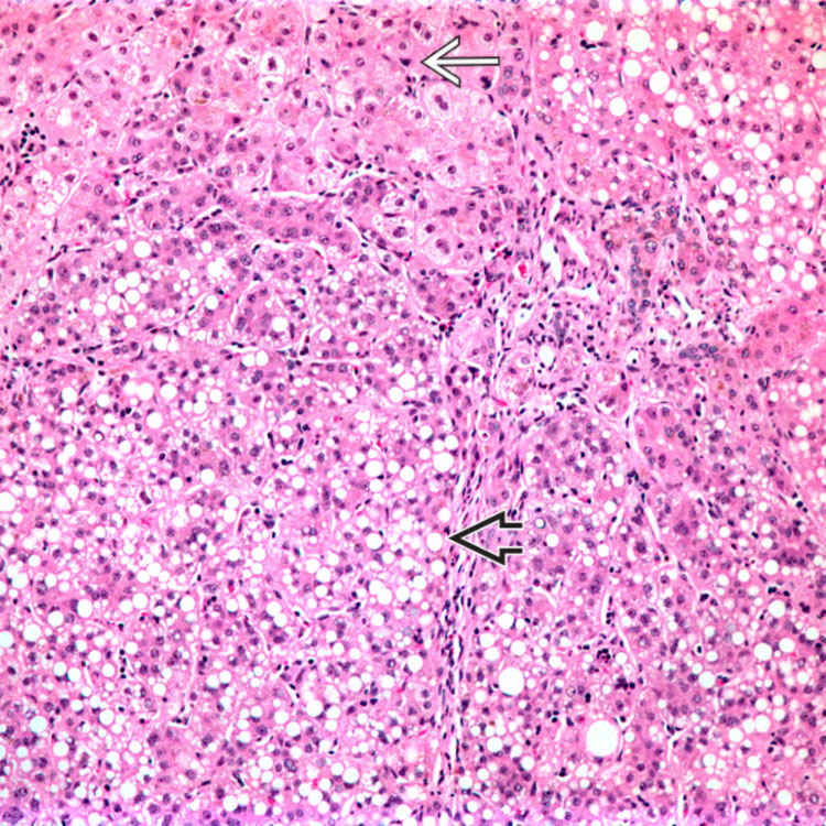
A dysplastic nodule
 in a patient with tyrosinemia displays both small cell change and widened plate architecture. Note the adjacent normal liver
  . (Courtesy M. J. Finegold, MD.)
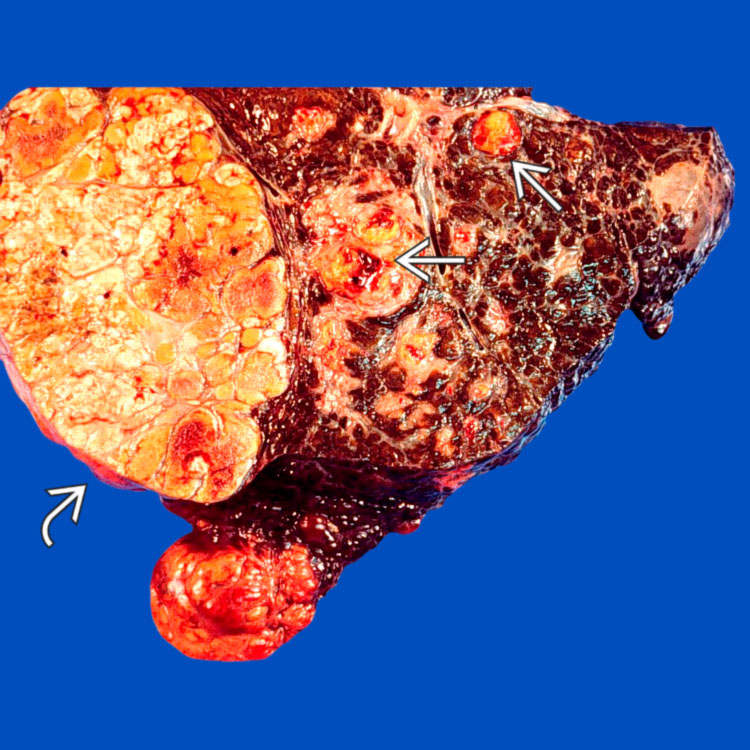
Gross photograph of an explanted cirrhotic liver from a 1.5 year old depicts a 7.5-cm multinodular, yellow mass
 as well as smaller nodules
  . (Courtesy M. J. Finegold, MD.)


ETIOLOGY/PATHOGENESIS
Molecular Basis
• Fumarylacetoacetate hydrolase deficiency due to 15q23-q25 gene mutations


Stay updated, free articles. Join our Telegram channel
Full access? Get Clinical Tree
