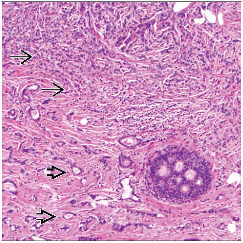
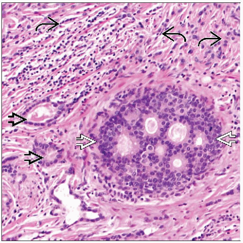
Tubulolobular Carcinoma
Key Facts
Terminology
Tubulolobular carcinoma (TLC)
Not a synonym for “carcinomas with ductal and lobular features”
Etiology/Pathogenesis
Low-grade invasive carcinoma with tubule formation and single cell lobular pattern
Initially described as tubular variant of lobular carcinoma
However, expression of E-cadherin supports classification as variant of ductal carcinoma
Reason for “lobular” appearance in presence of cell adhesion molecules has not been explained
Clinical Issues
1-2% of carcinomas
Microscopic Pathology
Admixture of round tubules and single cells infiltrating in single files
Ancillary Tests
Almost all cancers are positive for ER and PR and negative for HER2
Positive for E-cadherin and catenins
Top Differential Diagnoses
Tubular carcinoma
Invasive lobular carcinoma
Invasive carcinoma with ductal and lobular features
TERMINOLOGY
Abbreviations
Tubulolobular carcinoma (TLC)
Definitions
Morphologically distinct type of mammary carcinoma with 2 components consisting of minute well-formed tubules and single discohesive cells
TLC is not a synonym for carcinoma with ductal and lobular features
ETIOLOGY/PATHOGENESIS
Morphologic and Phenotypic Studies of TLC
Characterized by having both tubular (“ductal”) and single cell (“lobular”) morphologic components
Initially described as tubular variant of lobular carcinoma
Later studies demonstrated that TLC expresses E-cadherin and other cell adhesion molecules
Supports classification of TLC as variant of ductal carcinoma with lobular growth pattern
3-dimensional modeling of TLC and tubular carcinoma (TC)
Both tumor types consist of glandular structures connected by slender cords of single cells
Glands of TLC are connected by longer strands of single cells
TC has short strands of single cells that are less apparent in 2-dimensional sections
May account for appearance of tubular and lobular growth patterns in histologic sections
Morphologic appearance of loss of cohesion in presence of cell adhesion molecules is unclear
Catenin expression is normal in most TLCs
CLINICAL ISSUES
Epidemiology
Incidence
Rare; 1-2% of invasive breast carcinomas
Age
Similar to carcinomas of no special type; may be more common in older women
Gender
TLC has been reported in both males and females
Presentation
Usually presents with palpable mass
Less commonly found as a mass on mammographic screening
Prognosis
Intermediate prognosis between pure TC and invasive lobular carcinoma (ILC)
IMAGE FINDINGS
General Features
Location
Multifocality is present in ˜ 30% of cases
Positive lymph nodes are found in ˜ 60% of multifocal cases and ˜ 30% of cases with single focus
Size
Palpable mass present in 85% of patients
Mean size: 1.7 cm
Mammographic Findings
Most typical mammographic finding is irregular mass
May also show asymmetric focal density or architectural distortion
Ultrasonographic Findings
Mass with angulated, spiculated, or microlobulated borders
May have posterior acoustic shadowing or normal acoustic transmission
Stay updated, free articles. Join our Telegram channel

Full access? Get Clinical Tree


