Atypical Vascular Lesions of Skin
Key Facts
Terminology
Proliferation of endothelial-like cells within dermis of skin several years after exposure to therapeutic radiation treatment
Etiology/Pathogenesis
These lesions are only seen in clinical setting of prior radiation treatment
Clinical Issues
Lesions appear as fluid-filled vesicles or soft flesh-colored papules of skin
Complete excision of lesion is generally required for diagnosis and treatment
Majority of cases do not recur or develop malignancy after complete excision
Rare patients may develop angiosarcomas
Microscopic Pathology
Lesions consist of complex anastomosing vascular channels in dermis
Subcutaneous tissue or breast parenchyma is generally not involved
2 types
Lymphatic type (most common): Vessels are empty or filled with clear fluid
Vascular type (less common): Vessels are filled with blood
Top Differential Diagnoses
Angiosarcoma
Cutaneous hemangiomas
Acquired progressive lymphangioma
Reactive angioendotheliosis
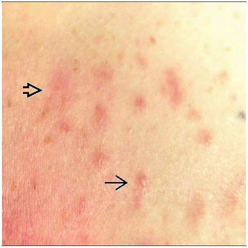 The majority of atypical vascular lesions (AVLs) present as small raised brown papules in the radiation field  . The 2nd most common appearance is as an ill-defined erythematous patch . The 2nd most common appearance is as an ill-defined erythematous patch  . . |
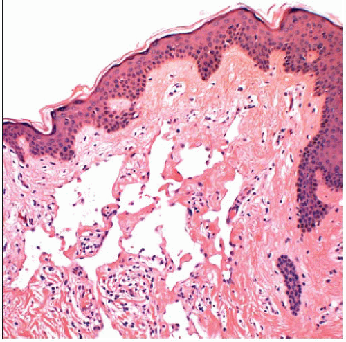 AVLs consist of anastomosing vascular channels within the dermis. The spaces usually appear empty (appearing as flesh-colored papules) but may be filled with blood (appearing as erythematous plaques). |
TERMINOLOGY
Abbreviations
Atypical vascular lesion (AVL)
Synonyms
Benign lymphangiomatous papules
Lymphangioma circumscriptum
Benign lymphangioendothelioma
Acquired progressive lymphangioma
Acquired lymphangiectasis
Definitions
Proliferation of endothelial-like cells within dermis of skin several years after exposure to therapeutic radiation treatment
ETIOLOGY/PATHOGENESIS
Etiology
These lesions are only seen in clinical setting of prior radiation treatment for carcinoma
Young women treated with radiation therapy for Hodgkin disease are at increased risk for breast carcinoma
AVLs have not been reported following radiation for Hodgkin disease
Reason why Hodgkin disease patients are not affected is not known
May be related to age at exposure and dosage
Radiation may damage cells lining lymphatics and small blood vessels
Radiation damage may cause abnormal proliferation
Typical time from radiation exposure to diagnosis of AVL: 3-4 years
CLINICAL ISSUES
Epidemiology
Gender
Only women have been reported to be affected
Presentation
2 types of AVLs
Lymphatic type (2/3)
Present as soft, flesh-colored (or pink to brown) papules, fluid-filled vesicles, or plaques
Vascular type (1/3)
Present as erythematous plaques or nodules
Gross appearance can closely mimic angiosarcoma
Patients may have lesions of both types
Treatment
Complete excision of lesion is generally required for diagnosis and treatment
In some cases, diagnostic features of angiosarcoma are apparent after complete excision
Benign-appearing lesions may progress to angiosarcoma over period of years
Complete excision may reduce likelihood of this
Prognosis
Majority of cases do not recur or develop malignancy after complete excision
However, 10-20% of patients experience recurrence at same site with AVL or develop new lesions
Rare patients progress to cutaneous angiosarcomas
It may be difficult to determine if angiosarcoma arose from AVL or is independent lesion
MACROSCOPIC FEATURES
General Features
Sections to Be Submitted
Stay updated, free articles. Join our Telegram channel

Full access? Get Clinical Tree


