Sarcomas
Key Facts
Terminology
Malignant mesenchymal neoplasm derived from connective tissue elements of the breast
Etiology/Pathogenesis
Mammary sarcoma can be de novo or secondary to prior treatment for carcinoma
Clinical Issues
Most common presentation: Palpable mass
Prognosis depends on histologic type of tumor, grade, and stage at presentation
May show aggressive behavior with local/systemic recurrence
Metastases to lungs, bone marrow, and liver most common
Metastases to lymph nodes are rare
Tumor size is significantly associated with survival
Microscopic Pathology
Virtually any type of sarcoma described elsewhere may occur rarely as primary lesion in the breast
Liposarcoma, leiomyosarcoma, osteosarcoma, and pleomorphic spindle cell sarcoma most common
Top Differential Diagnoses
Metaplastic carcinoma
Can show prominent sarcomatous differentiation
Immunostains for cytokeratin important to help confirm diagnosis
Phyllodes tumor of breast
May demonstrate extensive areas of overt sarcomatous overgrowth of stromal component
Look for biphasic pattern and epithelial component
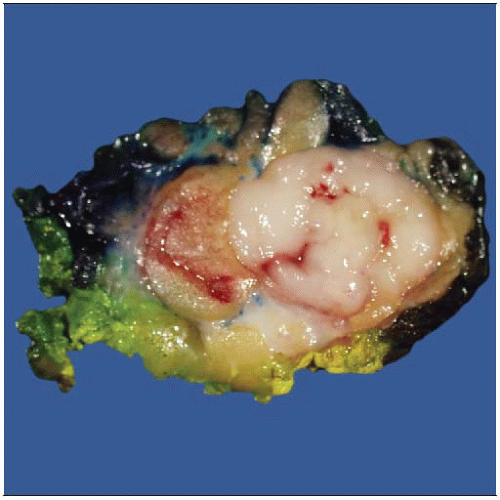 A 19-year-old female patient presented with a circumscribed palpable mass lesion thought to be a fibroadenoma based on clinical exam and imaging. Needle biopsy showed an undifferentiated malignancy. |
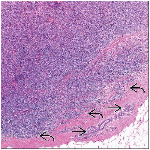 Section shows a high-grade spindle cell neoplasm with pushing borders  , compressing adjacent breast tissue , compressing adjacent breast tissue  . Further work-up showed this tumor to be a rhabdomyosarcoma. Staging studies were negative. . Further work-up showed this tumor to be a rhabdomyosarcoma. Staging studies were negative. |
TERMINOLOGY
Definitions
Malignant mesenchymal neoplasm derived from connective tissue elements of the breast
Sarcomas of the breast are rare
Sarcomatous-appearing tumors more likely to be metaplastic carcinoma or malignant phyllodes
Mammary sarcoma should be diagnosis of exclusion
Sarcoma involving breast tissue can be broadly divided into 3 categories
Idiopathic (de novo) sporadic cases (primary)
Post therapy (secondary)
Metastatic
Any sarcoma that occurs elsewhere can occur in the beast as a primary tumor
Angiosarcoma is most common primary sarcoma followed by liposarcoma
ETIOLOGY/PATHOGENESIS
Environmental Exposure
Etiology of most soft tissue sarcomas remains unknown
Primary mammary sarcoma of the breast can be de novo or secondary to prior treatment for carcinoma
Sporadic sarcomas tend to occur in younger age group
Sarcomas associated with prior use of breast external beam radiation therapy
Most common: Angiosarcoma, malignant fibrous histiocytoma, fibrosarcoma
Sarcomas association with chronic lymphedema that occurs after surgery with or without radiation treatment
Stewart-Treves syndrome
Lymphedema-associated lymphangiosarcoma
CLINICAL ISSUES
Epidemiology
Incidence
Mammary sarcomas: < 0.1% of breast malignancies
Primary sarcomas (other than angiosarcoma) are exceedingly rare
Annual incidence of breast sarcomas: 4.6 cases per 1,000,000 women
Presentation
Most common presentation: Palpable mass
Often rapidly enlarging
Natural History
Based on histologic type of tumor, grade, and stage at presentation
May show aggressive behavior with high likelihood for local/systemic recurrence
Hematogenous dissemination
Metastases to lungs, bone marrow, and liver are most common
Metastases to axillary lymph node are exceedingly rare
Treatment
Surgical approaches
Surgical excision with clean margins
Total mastectomy is most common
Smaller lesion may be treated with breast-conserving therapy
Axillary dissection is not indicated given the rarity of lymph node involvement
Adjuvant therapy
Any role for adjuvant chemotherapy &&/or radiation therapy is unclear
Prognosis
Primary mammary sarcomas have prognosis similar to that of their soft tissue counterparts
Based on histologic type, grade, and stage of tumor
Tumor size is significantly associated with overall survival (risk ratio = 1.3 per 1 cm increase)
Tumor grade is prognostically significant in some but not all reports
Post-Irradiation Sarcoma
Radiation therapy (RT) is important in adjuvant treatment of breast cancer
Complications include development of post-irradiation malignancy
Criteria for defining a post-irradiation sarcoma
Histological confirmation of sarcoma
Prior history of RT
Latency periods of several years until development of sarcoma
Development of sarcoma within previously irradiated field
Risk for development of post-irradiation sarcoma
0.03-0.8% with long-term follow-up (15 years)
IMAGE FINDINGS
Mammographic Findings
Lobulated or ill-defined mass
Architectural distortion
Ultrasonographic Findings
Circumscribed or spiculated mass
Ultrasound is generally better for delineating size of lesion
MACROSCOPIC FEATURES
Size
Superficial lesions tend to be smaller than those located deep in the breast
Larger lesions may show areas of hemorrhage and necrosis
Mean: 5.7 cm (range: 0.3-12.0 cm)
MICROSCOPIC PATHOLOGY
Histologic Features
Virtually any type of sarcoma described elsewhere may occur rarely as primary lesion in the breast
Leiomyosarcoma (LMS)
May arise from smooth muscle of nipple-areolar complex or vasculature
Morphologic and immunohistochemical findings are similar to LMS arising in other sites
Intersecting bundles of spindle cells with eosinophilic cytoplasm
Elongated nuclei with blunt ends and varying degrees of pleomorphism and mitotic activity
Liposarcoma (LS)
All grades of LS have been reported arising in the breast as a primary tumor
Stay updated, free articles. Join our Telegram channel

Full access? Get Clinical Tree


