Thrombosis, embolism and infarction

3 Thrombosis, embolism and infarction


THROMBOSIS


A thrombus is defined as a solid mass formed in the living circulation from the components of the streaming blood. This serves to distinguish it from a clot which may form:


Thrombosis (the formation of thrombus) is a well-ordered series of events involving the blood platelets and the clotting cascade. Platelets adhere to areas of endothelial damage and if the stimulus is strong enough will go on to platelet activation with shape change and release of a number of substances which enhance the process of thrombosis at the same time as aggregating together.


STAGES IN THE DEVELOPMENT OF THROMBOSIS


Thrombus may form in the heart, arteries, veins, or capillaries. The first stage involves platelets sticking to the damaged endothelium, and then a dense layer of fibrin and leucocytes adhere to the surface of the platelet. Blood clot (fibrin and red cells) develops on this layer of leucocytes and platelets, and then a secondary layer of platelets collects on the surface of the blood clot. The gradual extension of thrombosis leads to a propagated or consecutive thrombus. Organization then begins with adherence to the wall of the vessel as mural thrombus. A second stage develops with a further batch of platelets laid down over the initial aggregate and then a further layer of blood clot. In this way alternate layers of platelets and blood clot form a laminar arrangement. This causes a differential contraction of platelets and fibrin and gives a rippled appearance reminiscent of rippling of the sand on a beach. This has also been described as having a coralline appearance. The ridges on the surface of the thrombi are known as the lines of Zahn after the pathologist who first described them. Further development depends on whether the endothelium is healthy and on the rate of blood flow. Thus in an artery with thrombosis secondary to atherosclerosis, thrombosis may extend to the next branch after the endothelium becomes healthy again, assuming that there are collaterals with a reasonable blood flow. In veins, where the process tends to start in the pocket just above the valve, a number of things may happen: the process may end and the thrombus become covered with new endothelial cells; alternatively it may continue until a segment of vein is occluded. There is then a stagnant column of blood until the next tributary, and this stagnant column tends to coagulate, forming propagated thrombus. If the blood flow is reduced, the propagation may continue extensively. It may adhere to the sidewall of the veins in places or it may be largely free, simply attached to the site of origin. This latter type of thrombus can become dislodged relatively easily, forming a pulmonary embolism.


CAUSES OF THROMBOSIS


Several factors contribute to thrombus formation and these are usually grouped together under three headings (Virchows triad). The factors in Virchow’s triad are:


Not all these factors need to be present at the same time; some will be dominant in one clinical situation, whilst others will predominate in another. For example, venous thrombosis is commonly due to alterations in blood flow, while arterial thrombosis is more commonly due to vessel wall changes of atheroma, which does not occur in veins.


Damage to the vessel wall


Alterations in blood flow


The normal laminar flow may change to a turbulent pattern. This may happen with:


Alterations in blood flow are critical in the venous system since pressure is much lower and the normal rate of flow is much slower than in the arteries. As pressure is so much lower in the venous system and the vein walls are so much thinner than the walls of arteries of the same calibre, use is made of the pumping action of the surrounding muscle groups to aid return of blood to the heart. Consequently any decrease in muscle activity deprives venous blood of this added action and relative stasis occurs. Thus venous thrombosis becomes more likely in the veins of immobile subjects. The elderly are particularly at risk since they often have a degree of venous impairment or relative cardiac failure. One of the commonest deficiencies of the elderly venous system is impairment of the function of venous valves, and thrombosis is often seen to begin at the site of valves where, even under normal circumstances, some degree of turbulence is to be expected. For this reason it is particularly important to promote muscle contraction in the legs of the elderly in the postsurgical period. Another cause of relative immobility is long aeroplane journeys where immobility is combined with some degree of dehydration often aggravated by alcohol consumption.


Alterations in the constituents of the blood


Alterations which may occur include:


These may be:


FATE OF THROMBI


Thrombi may (Fig. 3.1):


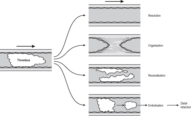
It is not clear what factors determine which of these fates a thrombus will suffer, although size may be a factor. Small thrombi are being formed and resolved constantly, and some degree of disturbance of blood flow is probably required to tip the scales and cause a thrombus to organise. Certainly a larger thrombus will cause turbulence and/or inflammation and make it likely that further thrombosis will occur on its surface, causing the thrombus to lengthen, a process known as propagation. Resolution means that the clot is completely dissolved by processes of thrombolysis. In the clinical setting this is achieved by the use of thrombolytic enzymes, e.g. plasminogen activator or urokinase, but these have to be delivered onto the clot more or less directly, otherwise they diffuse through the blood stream and may become so dilute that they are ineffectual. Current therapies involve substances that act directly or indirectly on plasminogen activators. Compounds such as aspirin and heparin help prevent further thrombus formation but do not help in lysis of an established thrombus. If the thrombus is not completely removed then the residue undergoes organisation.


Organisation is the process by which the thrombus is converted to a scar and eventually covered by endothelial cells. Intravascular scarring is essentially similar to those processes involved in the production of scars from thrombi in wound healing generally (Chapter 1). The main difference between intravascular granulation tissue and a thrombus is that with a thrombus the vascular phase of granulation tissue is prolonged and, if the thrombus does not resolve completely, the capillaries fuse together, resulting in one or several new vessels passing through the scar. This process is called recanalisation and in some cases may result in one or more functional vascular channels.


Thromboembolism is embolisation of a thrombus and should be distinguished from emboli of other materials since the clinical setting is different, as is the treatment. The effects of thromboemboli depend upon where the embolus settles, which in turn depends upon where the thrombus forms and what size the embolus is. Emboli arising from thrombi in veins will all go to the lungs (unless there is an abnormal connection between right and left heart). They will generally not arrest early in the circulation since the veins increase in diameter with the direction of blood flow as they approach the lungs, and only then do they start to turn into progressively smaller vessels of the lung bed. Arterial emboli will arrest in the artery with the smallest calibre which they can enter, and this will always be more peripheral than their origin because arterial size decreases in the direction of blood flow.


Dec 12, 2016 | Posted by in GENERAL & FAMILY MEDICINE | Comments Off on Thrombosis, embolism and infarction

Full access? Get Clinical Tree

Get Clinical Tree app for offline access