The trunk and neck

Part 4 The trunk and neck


Contents


Introduction


One of the major features distinguishing human beings from other animals is their bipedal posture and gait. As the hindlimbs progressively took over the locomotor function, the vertebral column assumed a new role. No longer is it held horizontally (Fig. 4.1A), where it is under compression, but it has become a vertical weight-bearing rod, held erect by ligaments and muscles (Fig. 4.1B). This change in function of the vertebral column has been accompanied by changes in its form, as well as by changes in its relationship to the skull and pelvic girdle.


Major differences in the form of the vertebral column are evident among quadrupeds, where it depends mainly on the distribution of mass in the animal. The so-called centre of gravity moves forward or backwards along the vertebral column in relation to there being more weight in the head and forelimbs or hindlimbs and tail respectively. In the evolution of the primates, the centre of gravity moved backwards towards the hindlimbs because both the length and musculature of the hindlimbs increased, in addition to the enlargement of the tail. The first of these changes gave additional power to leaping and grasping, while the latter provided balance when jumping. An important factor in locomotion is that the forward propelling force should pass through the centre of gravity; otherwise the body tends to rotate about the centre of gravity.


The early changes in body form during primate evolution made possible the sitting position, thereby freeing the forelimbs for manipulative activities. It also made possible the later stages of human evolution with the adoption of a bipedal gait. Although the human tail has been lost, the low position of the body’s centre of gravity has been maintained because of further development of the hindlimbs, a slightly reduced muscle mass in the forelimbs, and changes in the form and position of the trunk and abdomen.


A prerequisite for efficient arboreal locomotion was the evolution of an extremely flexible vertebral column. The increased range of flexion of the vertebral column, together with that of the lower limbs, gave additional propulsion in jumping as well as the ability to absorb the shock of impact on landing. However, in brachiating primates some flexibility of the vertebral column is lost due to it not having such an important role in locomotion. It seems that human beings could have evolved from a brachiating primate because of the relative stiffness of the vertebral column as well as other morphological features. In addition, four other fundamental changes have occurred in both the vertebral column and the thorax during human evolution, all of which are adaptations to a fully erect posture.


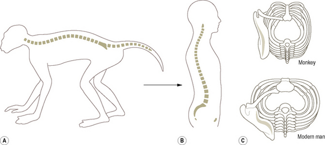
First, the vertebral bodies increase in size towards the lumbar region (Fig. 4.1B) because the compression forces along the trunk are no longer constant but increase progressively from above downwards. The proportions of the vertebral bodies also change from above downwards. Second, the spinous processes are more or less equally developed along the length of the vertebral column because the tendency to bend is no longer restricted to the limb girdles (the points of support), but is more evenly distributed. This is perhaps most clearly seen in the cervical region because the human head is more or less balanced on the vertebral column. Thirdly, the stiffening effect of the sternum and abdominal muscles on the vertebral column has become less important. The sternum has come to lie nearer the vertebral column so that the thorax is wider and less deep (Fig. 4.1C). This has resulted in a less abrupt change in direction of the ribs at their angles. Finally, the increased weight transmission through the pelvis and legs has brought about an enlargement of the sacrum, so that the human sacrum is usually composed of five fused segments. It is also relatively wide and more convex on its pelvic surface.


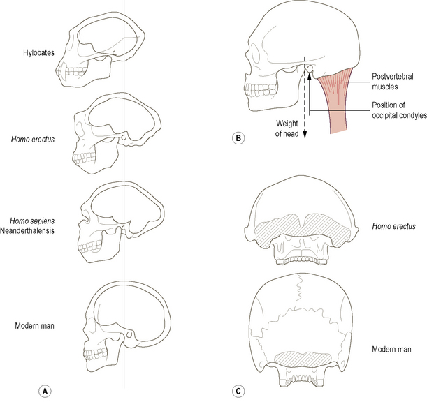
Concomitant with the changes in the vertebral column and trunk, the relation and form of the skull and its associated musculature have also changed dramatically. In quadrupedal animals, the head is supported by the postvertebral (nuchal) muscles and ligaments (these being under tension), which produce compression of the cervical vertebrae. As a more erect posture evolved the strength required in the postvertebral muscles was reduced because more of the weight of the head was carried directly by the vertebrae. This was accompanied by a reduction in the muzzle and an enlargement of the brain (Fig. 4.2A) causing the centre of gravity of the head to move more nearly over the point of support (the occipital condyles). It would be undesirable to have the head perfectly balanced on the occipital condyles because humans possess no powerful prevertebral muscles to support the skull from the front. Consequently, the centre of gravity projection falls just anterior to the occipital condyles, being balanced by the postvertebral muscles (Fig. 4.2B). That the postvertebral muscles have been reduced in importance is evident from the relatively small area of their attachment to the skull in humans compared with other primates (Fig. 4.2C).


In human beings the tail is reduced to between three and five fused coccygeal vertebrae, which curve ventrally and help form the pelvic cavity. Ligaments running from the coccyx to the ischium play an important role in maintaining this relationship, and in so doing contribute to the function of support of the abdominal and pelvic viscera. The abdominal viscera are carried in a sac-like cavity supported behind by the vertebral column, below by the pelvis and anterolaterally by the abdominal muscles (rectus abdominis, the external and internal abdominal obliques and transversus abdominis).


Although the vertebral column has changed in form and orientation during human evolution, it still has to fulfill essentially the same functional requirements as in quadrupedal animals:


The upright posture and independent functioning of the human upper limbs have greatly increased the dynamic demands made on the vertebral column. Nevertheless, the adaptation has been reasonably, although not altogether, successful such that the vertebral column has become a complicated and delicate mechanical unit. That the transition from quadrupedal to bipedal has not been entirely successful is witnessed by the fact that low back pain and its associated problems take a heavy toll.


The vertebral column comprises a series of mobile segments held together by ligaments and muscles, each separated from adjacent segments by an intervertebral disc. There are usually 33 bony segments, of which only 24 present as separate bones: the lower nine are fused, five forming the sacrum and four the coccyx. The 24 presacral vertebrae are designated cervical, thoracic and lumbar according to their features and position within the trunk: there are seven cervical, twelve thoracic and five lumbar vertebrae (Fig. 4.3). The length of the vertebral column is between 72 and 75 cm for the majority of individuals, of which approximately one-quarter is accounted for by the intervertebral discs. About 40% of an individual’s height is due to the length of the vertebral column. Variations in height between individuals mainly reflect differences in lower limb length rather than differences in vertebral column length. However, diurnal variations in height, up to 2 cm between early morning and late evening, are due to compression and loss of thickness of the individual intervertebral discs. (It is as well to remember this when charting the change in height of an individual.) Loss of height in elderly individuals is associated with thinning of the discs as a result of age-related changes within them.


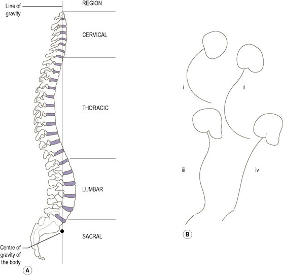
The adult vertebral column has four curvatures (Fig. 4.3A), an anterior convexity in the cervical and lumbar regions, and an anterior concavity in the thoracic and sacrococcygeal regions. Both the cervical and lumbar curvatures are acquired, in the sense that they are not present in early fetal development. Until late in fetal development, the vertebral column shows a single curvature concave anteriorly (Fig. 4.3B(i)). Late in fetal life the secondary cervical curvature (Fig. 4.3B(ii)) begins to appear, becoming more accentuated between 6 and 12 weeks after birth as the infant begins to hold up its head to enlarge its visual environment. The secondary lumbar curvature (Fig. 4.3B(iii)) appears when the child begins to sit up at around 6 months, becoming more marked with standing and the onset of walking. It is the extension of the hip that accompanies standing and walking which tilts the pelvis forwards so that the axis of the pelvic cavity is no longer in line with that of the abdominal cavity. The lumbar curvature develops in order to keep the trunk erect when standing. The lumbar curvature is not fully developed until after the age of 2, when a more or less adult pattern of walking is established. Sadly, in old age the vertebral column tends to assume a gentle C-shaped curve (Fig. 4.3B(iv)) reminiscent of the early fetal curve. The reason being that the shape of the vertebral column is largely determined by the intervertebral discs, and to a much lesser extent by the vertebrae themselves. Consequently, as the discs degenerate and thin with increasing age the secondary curvatures gradually disappear.


The relatively shallow cervical curvature begins in the dens of the axis and ends at the level of the second thoracic vertebra. It can be reduced or obliterated by bending the head forwards. The permanent thoracic curve is due in part to the shape of the thoracic vertebral bodies, the posterior height being greater than that anteriorly. This curvature ends at the 12th thoracic vertebra. An increase in the thoracic curvature is known as kyphosis. The lumbar curvature, which tends to be deeper and more prominent in women, ends at the lumbosacral junction. Changes in the orientation of the pelvis (pelvic tilt) are accompanied by changes in the lumbar curvature in an attempt to keep the trunk upright. The wearing of high-heeled shoes throws the pelvis forwards resulting in an increase in the lumbar curvature. A similar situation arises during pregnancy in an attempt to move the centre of gravity backwards and so prevent overbalancing. An increase in lumbar curvature is known as a lordosis, although the normal lumbar curve is often referred to as a lumbar lordosis. The curvature of the sacrum is permanent because of its fused constituent parts. It should be remembered that the lumbosacral angle is not a part of the vertebral curvatures. However, because it is the region where the mobile and immobile parts of the vertebral column meet, the structures associated with it, e.g. the intervening intervertebral disc and ligaments, are put under considerable stress.


The normal curvatures of the vertebral column make it a flexible support, imparting resilience to axial compressive forces which are absorbed by the giving way and recovery of the various curves.


Seen from the front, the vertebral column appears almost straight and symmetrical with perhaps a very slight right thoracic curve, probably due to the presence of the arch of the aorta. A large lateral curvature is abnormal and is known as a scoliosis. Scoliosis also involves rotation of the vertebral column so that the spinous processes of the vertebrae turn towards the concavity of the curvature. Compensatory curves in the reverse direction occur in order to keep the head facing forwards and over the feet. The condition is extremely complex, often appearing in childhood during periods of increased growth (i.e. 6–8 years, and during early puberty).


The vertebral curves pass in front of and behind the line of gravity along which the weight of the head, upper limbs and trunk is projected to the lower limbs. This line is said to pass progressively through the dens, the bodies of the second and 12th thoracic vertebrae and the promontory of the sacrum (Fig. 4.3), with the centre of gravity of the body located just in front of the sacral promontory. However, it must be remembered that the line of gravity is not constant; it is continually changing, both as the body moves and also when standing still. Nevertheless, it is a useful concept in reminding us of the natural balance and beauty of the body. By visualizing changes in the projection of this line, it may be possible to determine the structures put under increased strain in certain postures and pathologies.


Embryological development of the vertebrae


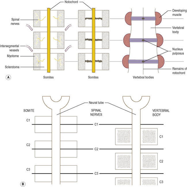
Soon after the formation of each somite it becomes differentiated into the three parts, the ventromedial sclerotome, the medial myotome and the thin lateral dermatome. The sclerotomes come to surround the notochord, followed by the myotomes. Each vertebra is formed from the adjacent parts of two sclerotomes (Fig. 4.4), with the intervening part forming the outer part (annulus fibrosus) of the intervertebral disc. The nucleus pulposus of the disc is derived from the notochord. As there are eight cervical somites, there are also eight cervical nerves, similarly in the thoracic, lumbar and sacral regions. However, because each vertebra is formed from adjacent somites the cervical nerves come to lie above their correspondingly numbered vertebrae, with the eighth cervical nerve lying below the seventh cervical vertebra. The thoracic, lumbar and sacral spinal nerves all lie below their correspondingly numbered vertebrae.


The dermomyotome breaks up with cells moving both ventrally and dorsally. The original spinal nerve supplying the myotome divides into an anterior and posterior primary ramus supplying the ventral (the hypomere) and dorsal (the epimere) parts of the myotome respectively. The epimeres come to lie between the transverse and spinous processes, giving rise to the musculature of the trunk and neck. The hypomeres give rise to the prevertebral muscles (the scalenes, quadratus lumborum, psoas and piriformis) and the musculature of the thoracic and abdominal walls.


The occipital myotomes do not participate in the formation of the musculature of the trunk or neck. They give rise to the muscle mass of the tongue. That part of the paraxial mesoderm that does not become segmented eventually becomes incorporated into the branchial (pharyngeal) arches, each of which is innervated by a cranial nerve. The musculature derived from these arches forms the muscles of the face, those around the mandible and those of the larynx and pharynx.


The vertebral column


Extending from the base of the skull to the pelvis, the vertebral column consists of a series of irregularly shaped bones which increase in size from above downwards. The vertebrae are bound together by ligaments and have intervertebral discs between their bodies. In young children, 33 separate vertebrae can be identified; however, by the time adulthood is reached five have fused to form the sacrum and four to form the coccyx. Of the remaining 24, seven are found in the neck (cervical vertebrae), twelve articulate with the ribs (thoracic vertebrae) and five are found in the lower back (lumbar vertebrae). Within each group the vertebrae have similar features, some of which are distinctive, regarding shape and the orientation of their articular processes.


With the exception of the first and second cervical vertebrae, all vertebrae possess a large weight-bearing body anteriorly and vertebral arch posteriorly, which consists of a series of bony processes (Fig. 4.5A). The body varies in shape and size depending on its location in the vertebral column, but is roughly cylindrical with flattened upper and lower surfaces. On these surfaces, the markings of the attachment of the intervertebral disc around the periphery can be seen surrounding a roughened central area. The front and sides of the bodies are roughened, particularly at their upper and lower edges, and concave from top to bottom. The posterior surface is fairly smooth and has a large foramen for the passage of the basivertebral vein. The vertebral arch arises from the posterolateral aspect of the body, and with the body surrounds the vertebral foramen. In situ adjacent vertebral foramina, together with the intervertebral discs and the ligamenta flava, form the vertebral canal which houses the spinal cord and its various coverings.


The vertebral arch consists of two pedicles and two laminae. Each pedicle passes from the upper part of the posterolateral aspect of the vertebral body to join with the anterolateral extremity of the corresponding lamina. The laminae slope backwards to meet in the midline posteriorly, where they are continuous with the posterior projecting spinous process. Because the pedicles are not as deep as the vertebral bodies, the opening formed between adjacent pedicles (the intervertebral foramen) enables the spinal nerves and supporting blood vessels to leave or enter the vertebral canal. The intervertebral foramen is closed anteroinferiorly by the intervertebral disc, an important relation to note in some pathologies of the vertebral column and spinal nerves.


Arising from the junction of each pedicle and lamina are three processes: the transverse process projecting laterally, and the articular processes, one directed upwards and one directed downwards. In adjacent vertebrae, superior and inferior articular processes make contact with each other by small synovial joints – the zygapophyseal joints. It is the shape and orientation of the articular facets on these processes which largely determine the range and type of movement possible between adjacent vertebrae.


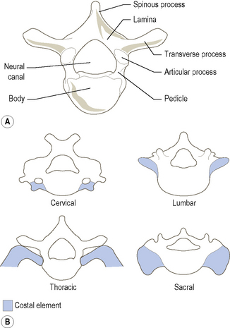
With a few exceptions, each part of a vertebra is potentially present in every other vertebra, the main features of which are shown in Fig. 4.5A. The body is the only part of a vertebra that is represented throughout the whole series of vertebrae; even so, that of the atlas has been displaced as the dens of the axis. The major difference between cervical, thoracic, lumbar and sacral vertebrae is related to the size and fate of their costal elements (Fig. 4.5B). In the cervical region, the medial end of the costal element fuses with the side of the vertebral body. The presence of the vertebral vessels lying anterior to the true transverse process prevents complete fusion of the costal element with the transverse process. Instead, a costotransverse lamella (bar) joins the two parts together laterally, leading to the formation of the foramen transversarium. Consequently, the anterior tubercle of the transverse process is the lateral end of the costal element, while the posterior tubercle represents the lateral end of the transverse process. The first and second cervical vertebrae have no anterior tubercles.


In the thoracic region, the costal element remains separate giving rise to the rib, which articulates directly with the body and transverse process of its corresponding vertebra.


In the lumbar region, the costal element has again been incorporated into the vertebra. So complete is its incorporation that apart from the root of the transverse process (lateral process) the remainder is composed of costal element. The true transverse process in the lumbar region gives rise to the accessory and superior processes as well as the lateral process.


Even in the sacrum, the costal elements have been incorporated and form the major part of the lateral masses.


Bones


A description of the sacral and coccygeal vertebrae can be found on pages 211–213.


Lumbar vertebrae


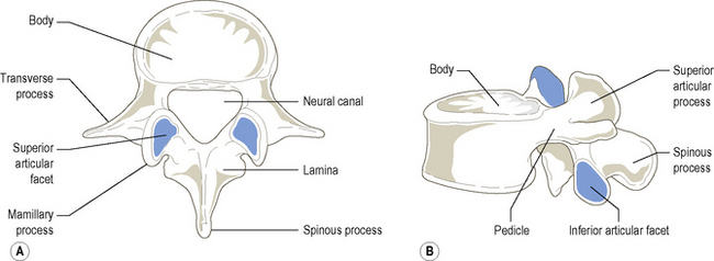
The five lumbar vertebrae are much stouter and stronger than those in either the thoracic or cervical regions (Fig. 4.6). They possess neither foramina transversaria nor articular facets for the ribs. Each has a large, kidney-shaped body with almost parallel upper and lower surfaces, except for L5 which is deeper anteriorly than posteriorly. The short strong pedicles pass almost directly backwards to join the narrow laminae which pass backwards and medially towards the spine. Adjacent laminae are widely separated from each other, leaving diamond-shaped spaces which contain the ligamenta flava.


The spinous processes of the lumbar vertebrae project almost horizontally backwards, being level with the lower half of the body. They are wider from above downwards with a thickened posterior edge. That of L5 is frequently rounded.


The articular processes project superiorly and inferiorly from the region where the pedicle joins the lamina. The articular facets on the superior process are concave transversely and flat vertically: they face posteromedially. On the posterior edge of the superior articular process is the rounded mamillary process. The inferior articular processes are set closer together than the superior and have facets which are reciprocally curved to face anterolaterally. The superior facets articulate with the inferior facets of the vertebra immediately above. The inferior articular processes of the fifth lumbar vertebra are more widely set apart and flatter than those of other lumbar vertebrae. Their articular facets face anterolaterally to meet the superior articular facets of the sacrum.


The triangular vertebral (neural) canal is larger than that in the thoracic region, but slightly smaller than in the cervical region.


With the exception of the fifth lumbar vertebra the transverse processes are short and thin, projecting laterally and slightly backwards from the sides of the vertebral body and base of the pedicles. The third is the longest, while the fourth and fifth are inclined upwards. The transverse processes of L5 are short and stout, and may be fused with the lateral part of the sacrum. From the root of each transverse process, a small tubercle (the accessory process) projects posteriorly. The root of the transverse process is known as the lateral tubercle.


Thoracic vertebrae


The distinctive feature of thoracic vertebrae is the presence of articular (costal) facets on the sides of the vertebral body for articulation with the heads of at least one pair of ribs (Fig. 4.7). The bodies of thoracic vertebrae are typically heart-shaped, when viewed from above, and on their sides bear articular facets. The bodies of the second to eighth vertebrae have large, almost complete oval facets near their upper borders, and smaller demifacets near their lower borders. Although similar in basic arrangement, the upper facet of the ninth thoracic vertebra is situated at the base of the pedicle. The body of T1 has a complete oval facet near its upper edge and a demifacet at its lower. The 10th, 11th and 12th vertebrae have single complete facets located at the junction of the body with the pedicle. Between T10 and T12 the facets gradually move from the upper to lower region of the junction (Fig. 4.7B).


The short pedicles project almost directly backwards from the upper posterior part of the body. They gradually become larger and stronger from above downwards. The laminae are inclined towards the midline, and although narrow from side to side they are deep so that they overlap one another from above.


The spinous processes are long and slope downwards, with those in the middle of the series being almost vertical. The upper and lower ones are shorter and generally less sloping. The 12th is almost horizontal, resembling a typical lumbar spine.


The long, thick, rounded transverse processes project laterally, backwards and slightly upwards from the junction of the pedicle with the lamina. They have an oval facet on the anterior surface near the tip which faces anterolaterally, for the tubercle of the corresponding rib. In the upper thoracic vertebrae these facets are concave but gradually become flatter from above downwards. The transverse processes of the 11th and 12th thoracic vertebrae have no facets. The transverse process of the 12th thoracic vertebra is very short and shows features similar to the lumbar vertebrae.


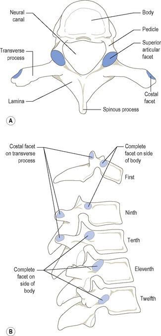
From just medial to the base of the transverse process, the articular processes project almost vertically, the superior upwards and the inferior downwards. The articular facets on the superior process are slightly concave transversely, being flat from above down, and face backwards but also slightly upwards and laterally. The facets on the shorter inferior articular processes are reciprocally curved and face in the opposite direction. The superior articular facets of one vertebra articulate with the inferior facets of the vertebra above. The joints so formed lie on the arc of a circle whose centre lies within or just anterior to the body of the vertebra. Consequently, rotation as well as flexion and extension are favoured in the thoracic region (p. 463).


The inferior articular processes of T12, although they project vertically, do not lie in the same general plane as the other thoracic articular processes. They are in fact lumbar in type; therefore they are markedly convex transversely and face anterolaterally.


The vertebral (neural) canal is smaller than that in either the cervical or the lumbar region, and is nearly circular in appearance.


Cervical vertebrae


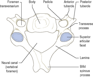
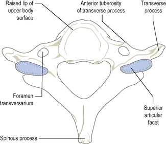
The characteristic feature of all cervical vertebrae is the presence of a foramen transversarium in each transverse process (Fig. 4.8). They tend to be small as they do not carry much weight. The third, fourth, fifth and sixth cervical vertebrae are sufficiently similar to be considered together. The body is relatively small, appearing kidney-shaped when viewed from above. Its superior surface projects upwards at the sides, while its inferior surface is correspondingly bevelled. Between the sides of adjacent surfaces of the bodies, each side of the intervertebral disc, are small synovial joints (uncovertebral joints, p. 451). The anterior surface is marked by the attachment of the anterior longitudinal ligament, while laterally the body is hollowed. The posterior surface of the body is flat.


The short pedicles pass laterally and backwards, which is why the vertebral canal in the cervical region is triangular and larger than elsewhere. The long narrow laminae pass posteromedially, joining together to form the spinous process. The short, bifid spinous process projects backwards from the centre of the vertebral arch.


The composite transverse processes are stout projections arising from the lateral side of the body and pedicle. Each transverse process ends in prominent anterior and posterior tubercles, joined by the costotransverse lamella. Within each process is a foramen transversarium, bound posteriorly by the pedicle, and anteriorly and laterally by the various parts of the transverse process. It transmits the vertebral artery and vein.


The large superior and inferior articular processes project from the articular mass, which is located at the junction of the pedicle and lamina on each side.


Each process bears an articular facet. The slightly convex superior facet faces upwards and backwards while the reciprocally concave inferior facet faces downwards and forwards. The facets become more vertical in the lower part of the cervical spine.


The seventh cervical vertebra


The seventh vertebra (Fig. 4.9), also known as the vertebra prominens, is noted for the length of its non-bifid spinous process. It is larger than the preceding cervical vertebra and exhibits similarities to thoracic vertebrae. The body is larger, the pedicles are directed more posteriorly than laterally, the inferior articular facets face more anteriorly than downwards, and the vertebral canal is generally smaller than that of other cervical vertebrae.


The transverse processes have a small foramen transversarium, which only transmits the vertebral vein. Occasionally the anterior costal element of the transverse process is much longer than the posterior part, giving the appearance of a rudimentary rib as it extends forwards towards the first rib as either a fibrous or bony strip. This may lead to pressure on the eighth cervical nerve root as it passes forwards over the first rib to take part in the brachial plexus.


The axis (C2)


The axis (C2), the second and strongest of the cervical vertebrae, has many of the features of a typical cervical vertebra (Fig. 4.10). It has, however, the separated body of the atlas fused with the superior surface of its body giving an upward tooth-like projection known as the odontoid process (dens). The dens is slightly pointed at its apex, where the apical ligament attaches it to the anterior margin of the foramen magnum. From the sloping sides of the apex, alar ligaments pass to tubercles on the medial sides of the occipital condyles. Its anterior surface has a small smooth facet, concave from above down and convex transversely for articulation with the facet on the back of the anterior arch of the atlas. Posteriorly is a smooth, constricted horizontal surface where the transverse ligament crosses. Above this constriction there is a slightly expanded area known as the head.


The large superior articular facets lie at the junction of the body and pedicle, and transmit the weight of the head to the body of the axis, leaving the dens free to rotate with respect to the atlas. The articular facets are slightly convex, resembling a segment of a dome, and face upwards and laterally. They allow the gliding forwards of one lateral mass while the other glides backwards when the atlas rotates on the axis. The slightly concave inferior facets face downwards and forwards and are situated just behind the transverse processes at the junction of the pedicles and the laminae on either side.


The transverse processes are small and rounded, projecting laterally from the sides of the vertebral body. As there are no anterior tubercles, the front of the foramen transversarium is closed by the costotransverse lamella.


The thick and strong laminae pass backwards and medially, joining together to form the strong stout spinous process which is usually bifid. The spinous process projects almost 1 cm further posteriorly than the posterior tubercle of C1 and covers the much thinner spinous process of C3.


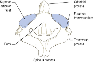
The inferior surface of the axis is very similar to that of a typical cervical vertebra (Fig. 4.8).
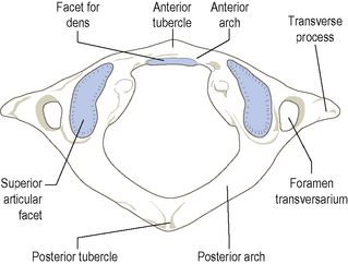


The atlas (C1)


This ring of bone bears very little resemblance to a cervical or for that matter any other vertebra (Fig. 4.11). It has no body or spine, but consists of slender anterior and posterior arches joined on each side by a lateral mass, which bears articular facets superiorly and inferiorly and a transverse process laterally. The concave superior facets articulate with the condylar processes of the occipital bone, while the concave inferior facets articulate with the second cervical vertebra. The inferior articular facets are segments of a sphere, thereby facilitating rotation between the atlas and axis. Each lateral mass is marked on its medial side by a small tubercle to which the transverse ligament of the atlas attaches.


Passing between the lateral masses anteriorly is the short flattened anterior arch, which has in the middle of its anterior surface the anterior tubercle and posteriorly a facet for articulation with the dens of the axis. The posterior arch, which represents the pedicles and laminae of typical vertebrae, is a long, curved bar of bone joining the lateral masses and the roots of the transverse processes. Posteriorly, the posterior tubercle represents the spinous process. On the superior surface of the posterior arch, running medially from the back of each lateral mass, is a shallow groove for the vertebral artery before it enters the foramen magnum. This groove may be converted into a foramen by cartilaginous or bony tissue.


The strong transverse processes are large and wide. They may also be bifid, even though they represent only the posterior tubercle of a typical cervical vertebra. The foramen transversarium, lying close to the lateral mass, transmits the vertebral vessels as well as sympathetic nerves.


Section summary


The vertebrae


The vertebral column


Ossification of the vertebrae


A typical vertebra ossifies in cartilage from three primary ossification centres and five secondary ossification centres.


The primary centres appear in the vertebral body and in each half of the vertebral arch. That for the centrum, the larger median part of the body, appears between the second and fifth month in utero, being present first in the lower thoracic region and then spreading sequentially up and down the column. The centres for the coccygeal vertebrae appear between birth and puberty, with ossification spreading without the formation of secondary centres. Because each centrum is usually ossified from two centres, anomalies may arise in that they may fail to unite and remain as two separate halves, or only one may ossify giving rise to a hemivertebra.


The primary centre in each half of the vertebral arch appears at the junction of the pedicle and lamina at 2 months in utero in the upper cervical region, spreading down to the sacrum by the fifth month. From each centre, ossification spreads into the lamina and pedicle where it extends into the centrum to complete the body, and into the root of the transverse process.


At birth, the centrum and each part of the vertebral arch are separated by cartilage. That between the centrum and each arch begins to ossify in the cervical region during the third year, extending to other regions by the seventh year. The laminae begin to unite soon after birth in the lumbar region, spreading to the cervical region by the second year. This process is not complete in the sacrum until the seventh to tenth year. Once the laminae have fused, ossification spreads into the root of the spinous process.


The multiple secondary centres for the upper and lower surfaces of the bodies appear during the ninth year. They fuse to form flat rings of bone around the periphery of these surfaces. Secondary centres appear in the tips of the transverse processes during the 18th year. Fusion of all of these secondary epiphyses with the rest of the vertebra begins at 18, for the bodies, and is complete by age 25.


The lumbar vertebrae also have secondary centres for the mamillary processes. In addition, the first lumbar vertebra may have separate primary centres for its transverse processes, which may remain separate to form a lumbar rib. In the fifth lumbar vertebra there may be two primary centres in each half of the vertebral arch, united by cartilage between the superior and inferior articular processes. Consequently, there is a temporary risk of separation between the two parts.


The upper two cervical vertebrae cannot be considered typical in their pattern of ossification and therefore are dealt with separately. In the atlas a primary centre appears during the second month in utero for each half of the vertebral arch and the associated lateral mass, the two halves usually unite during the third or fourth year. Occasionally, a secondary centre may appear in the intervening cartilage. At birth the anterior arch is cartilaginous, but an ossification centre appears by the end of the second year to fuse with the lateral masses, and includes the anterior part of the superior articular surface, during the seventh year. An epiphysis for each transverse process appears and unites with the rest of the bone during puberty.


In the axis, primary centres for each vertebral arch appear during the second month in utero, one for the lower part of the body and two more side by side for the dens and upper part of the body during the fifth month. Those for the dens fuse together 2 months later so that at birth the axis consists of four parts which unite between the third and sixth year. Secondary centres appear in the tip of the dens between 2 and 6 years to fuse with the dens by the age of 12, and for the lower surface of the body which fuses during puberty between 18 and 25. Occasional additional ossification centres may be present.


Bifid cervical spinous processes each have a secondary ossification centre, in addition to which the sixth and seventh cervical vertebrae may have separate primary ossification centres for their costal elements. If present, these usually fuse with the rest of the bone during the fifth year. However, there may be a tendency for that of the seventh to remain separate as a cervical rib.


With so many ossification processes and patterns occurring simultaneously it is not surprising, therefore, to find some variations in the total number of vertebrae present. This is usually due to a reduction, or rarely, an increase in the number of coccygeal vertebrae.


The number of cervical vertebrae is constant at seven. However, the number of thoracic vertebrae may be increased by the presence of ribs associated with L1. Similarly the number of lumbar vertebrae may be reduced as above, or by incorporation of L5 into the sacrum. Such sacralization may be partial or complete. The sacrum may gain or lose additional segments.


Many congenital conditions of the vertebral column are due to incomplete fusion of its constituent parts. Hemivertebrae can cause an abnormal lateral curvature (scoliosis) of the vertebral column. The laminae may fail to fuse or meet in any region, but most commonly in the lumbosacral region, giving rise to spina bifida. The spine, laminae and inferior articular processes of L4 and L5 may be joined to the rest of the vertebrae by cartilage and not completely fused. Under certain loading conditions this can lead to a separation whereby the vertebral body, most commonly that of L5, slides forwards. This condition is known as spondylolisthesis (see p. 284).


Palpation of the vertebral column


Unfortunately there is considerable variation in the location of bony points from one individual to another and even from side to side within the same individual. This is particularly apparent in the region of the trunk, where the length of the spines can vary considerably, be angled differently or occasionally even be absent. The best way of identifying vertebral spines is therefore to count downwards or upwards from known bony landmarks and cross check with other surface markings. Considerable time and practice is involved in developing palpation techniques, but once mastered it will prove to be an invaluable asset in the future.


The most obvious surface markings are the posteriorly directed spines. However, their palpation can vary considerably, due to the lordosis (convexity forwards) in the cervical and lumbar regions and the kyphosis (concavity forwards) in the thoracic and sacral regions of the vertebral column.


Lumbar region


This region of the vertebral column has a lordosis similar to that of the cervical region. Consequently, it is not easy to identify the spinous processes. With the subject lying prone, and with sufficient support under the abdomen to raise the lumbar region so that it is level, the whole lumbar region can be examined. The spinous processes of the lumbar vertebrae can be palpated in a central cleft down the midline. About 3 cm each side of the midline a small dimple can be seen on the posterior superior aspect of the buttock. This marks the location of the posterior superior iliac spine, which can be easily palpated and acts as an important landmark for the identification of other structures. From these spines the crests of the ilium can be traced upwards and forwards. The spinous process of L5 can be felt in a deep hollow, just above the sacrum, approximately 2 cm above a line drawn between the posterior superior iliac spines. From here the spinous process of L4 is easily recognizable above that of L5. With care, the small gaps between the spinous processes of L4 to T12 can be palpated, and each process identified. The centre of the spinous processes of each vertebra, unlike that in the thoracic region, lies at a level just below the centre of the body of its corresponding vertebra. On either side of the midline is a powerful column of muscle tissue running from the posterior part of the sacrum up towards the thoracic region.


On deep palpation, lateral to this bulk of muscle, small pointed tubercles can be felt, running down either side. These are the tips of the transverse processes, each one being located just above the level of the centre of its corresponding spinous process. Higher up, level with the spinous process of L1, the tip of the 12th rib can be palpated, being level with the ninth costal cartilage in front and lying in the transpyloric plane.


May 25, 2016 | Posted by in ANATOMY | Comments Off on The trunk and neck

Full access? Get Clinical Tree

Get Clinical Tree app for offline access