Teratoma
Steven S. Shen, MD, PhD
Jae Y. Ro, MD, PhD
Key Facts
Terminology
Tumors with somatic tissue of different germinal layers (ectoderm, mesoderm, or endoderm)
Clinical Issues
2nd most common childhood germ cell tumor in infants and young children
Pure teratoma rare in adult and is often present mixed with other germ cell tumor
Macroscopic Features
Often well-circumscribed, nodular and firm mass with heterogeneous cut surface with solid and cystic areas
Microscopic Pathology
Mature teratoma
Composed of mixture of elements of ectoderm, endoderm, and mesoderm
Most common components are different types of epithelium, cartilage, or nerve
Immature teratoma
Undifferentiated spindle cell component or primitive neuroectodermal tissue
Stromal overgrowth with foci of embryonal rhabdomyosarcoma, Wilms tumor-like elements, or angiosarcoma
Carcinomatous transformation with invasive growth
Top Differential Diagnoses
Primary or metastatic sarcoma
Mixed germ cell tumor
Metastatic carcinoma
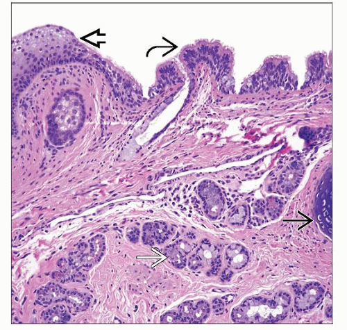 This mature teratoma shows squamous  and respiratory epithelium and respiratory epithelium  , seromucinous glands , seromucinous glands  , and cartilage , and cartilage  . Virtually all somatic tissue types may be seen in a teratoma. . Virtually all somatic tissue types may be seen in a teratoma. |
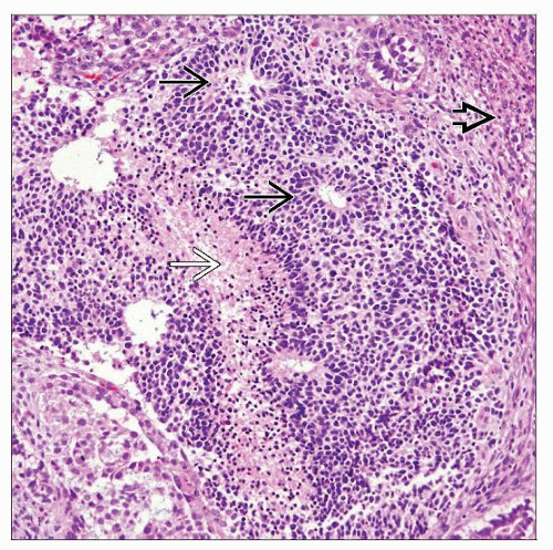 Immature teratoma shows neuroectodermal cells and neural tube-like structures  . Focal necrosis . Focal necrosis  and cellular spindle immature mesenchymal tissue and cellular spindle immature mesenchymal tissue  are also present. are also present. |
TERMINOLOGY
Synonyms
Mature teratoma, immature teratoma
Definitions
Tumors with > 1 somatic tissue of different germinal layers (ectoderm, mesoderm, or endoderm)
ETIOLOGY/PATHOGENESIS
Genetics
Pediatric teratomas are diploid
Adult teratomas are often aneuploid (hypotriploid)
CLINICAL ISSUES
Epidemiology
Incidence
Pure form constitutes 4-9% of all testicular tumors
2nd most common childhood germ cell tumor after yolk sac tumor in infants and young children
Pure teratoma in adults is extremely rare
Frequently mixed with other germ cell tumor types (approximately 50%)
Age
Occurs in 2 distinct age groups: Pediatric (< 4 years of age) and adults (2nd to 4th decades)
Presentation
Painless firm testicular mass
Treatment
For teratoma in prepubertal children, orchiectomy without lymph node dissection
For teratoma in adults, regardless of maturation, at least orchiectomy with close follow-up
Prognosis
Prepubertal teratomas are almost always benign
Adult teratomas are considered malignant because of relatively high recurrence or metastasis (22-37%)
IMAGE FINDINGS
General Features
Solid and cystic testicular mass by ultrasound
MACROSCOPIC FEATURES
General Features
Often well-circumscribed, nodular and firm mass with heterogeneous cut surface with solid and cystic areas
Cysts filled with clear, white, flaky, gelatinous or mucoid material
Mature tissue with hair, cartilage, bone, or teeth may be seen
Size
Variable
MICROSCOPIC PATHOLOGY
Histologic Features
Mature teratoma
Composed of mixture of elements of ectoderm, endoderm, and mesoderm
Ectoderm: Epidermis, neuronal tissue
Endoderm: Gastrointestinal or respiratory mucosa, other seromucous glands
Mesoderm: Bone, cartilage, muscle
Most common components are different types of epithelia, cartilage, or nerve
Respiratory and gastrointestinal epithelium, muscle, and cartilage are more commonly seen in testis than in ovary
Pancreatic, dental, renal, and thyroid tissue are less commonly seen in testis than in ovary
Immature teratoma
Primitive mesoderm: Undifferentiated spindle cell component (most common immature element in testis)
Primitive endoderm and primitive neuroectoderm (resembling neural tube and embryonic nervous system)
Blastomatous tissue (resembling blastema and embryonic tubules of developing lung or kidney), embryonic rhabdomyoblastic tissue
Teratoma with secondary malignant (somatic type) transformation
Sarcomatous transformation of teratoma: Foci of embryonal rhabdomyosarcoma, Wilms tumor-like element, or angiosarcoma
Carcinomatous elements in teratoma (such as squamous cell carcinoma, adenocarcinoma) with invasive growth
When histology of malignant component forms pure nodule of substantial size (> 1 field of 4x objective)
Cytologic Features
Highly variable and depends on tissue type and maturity
Predominant Pattern/Injury Type
Neoplastic
Predominant Cell/Compartment Type
Variable tumor cells from > 1 germ cell layer
ANCILLARY TESTS
Immunohistochemistry
Highly variable and depends on component of teratoma (rarely necessary in clinical practice)
Cytokeratin, CEA, and EMA/MUC1: Positive in epithelial tissue or carcinoma of teratomatous type
Vimentin: Positive in mesenchymal tissue
Germ cell markers: HCG (syncytiotrophoblastic cells), AFP (enteric and hepatoid tissue), PLAP (may be glandular tissue)
Other tissue specific markers for different type of tissues
DIFFERENTIAL DIAGNOSIS
Primary or Metastatic Sarcoma
Usually involves the paratesticular structures, more homogeneous population of pleomorphic spindle cells
Stay updated, free articles. Join our Telegram channel

Full access? Get Clinical Tree


