Syringomatous Adenoma of the Nipple
Key Facts
Terminology
Benign neoplasm morphologically similar to dermal syringoma
Clinical Issues
Presents as palpable, solitary, firm dermal mass in subareolar or nipple region
Management is complete local excision to achieve negative margins
Local recurrences in up to 30% of incompletely excised lesions
No risk for local recurrence if completely excised with negative margins
Does not metastasize
Image Findings
May be indistinguishable from carcinoma on mammography or ultrasonography
Microscopic Pathology
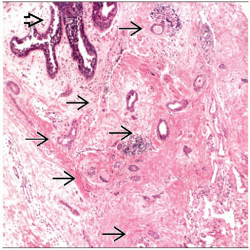
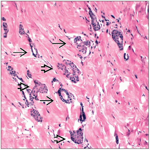
Infiltrative proliferation of eccrine duct-like structures
Squamous cell nests and glandular structures are present
Infiltration into smooth muscle bundles and nerves simulates malignant neoplasm
Cells have small uniform nuclei
Mitotic figures are not usually present
Overlying epidermis may be acanthotic
Top Differential Diagnoses
Tubular carcinoma
Low-grade adenosquamous carcinoma
TERMINOLOGY
Abbreviations
Syringomatous adenoma of the nipple (SAN)
Synonyms
Infiltrating syringomatous adenoma
Syringomatous tumor
Definitions
Benign, locally invasive neoplasm of probable eccrine duct origin that forms palpable mass in areolar dermis
ETIOLOGY/PATHOGENESIS
Cell of Origin
Most likely origin is intraepidermal eccrine sweat ducts of nipple
Morphologically identical to syringomas of skin in other parts of body that arise from eccrine ducts
CLINICAL ISSUES
Epidemiology
Incidence
Very rare
Age
Mean: 40 years (range: 11-76 years)
Gender
Almost all patients have been female
Only 1 reported case in a male
Presentation
Solitary firm mass in subareolar or nipple region
May be associated with
Pain or tenderness
Skin crusting or ulceration
Nipple discharge
Nipple retraction
Treatment
Surgical approaches
Should be completely excised with negative margins
Prognosis
Benign lesion
No reported cases of regional or distant metastasis
May recur locally if not completely excised (30% of cases with positive margins)
IMAGE FINDINGS
General Features
May be indistinguishable from carcinoma on mammography or ultrasonography
Mammographic Findings
Mass-forming lesion in subareolar region
Borders may be irregular
Calcifications may be present
Ultrasonographic Findings
Ill-defined mass with heterogeneous internal echoes
MACROSCOPIC FEATURES
General Features
Ill-defined, firm, white mass with minute cystic spaces in the dermis of the nipple and areola
Size
1-3 cm
Stay updated, free articles. Join our Telegram channel

Full access? Get Clinical Tree


