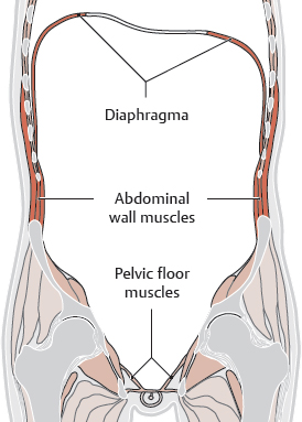
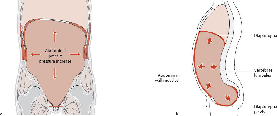
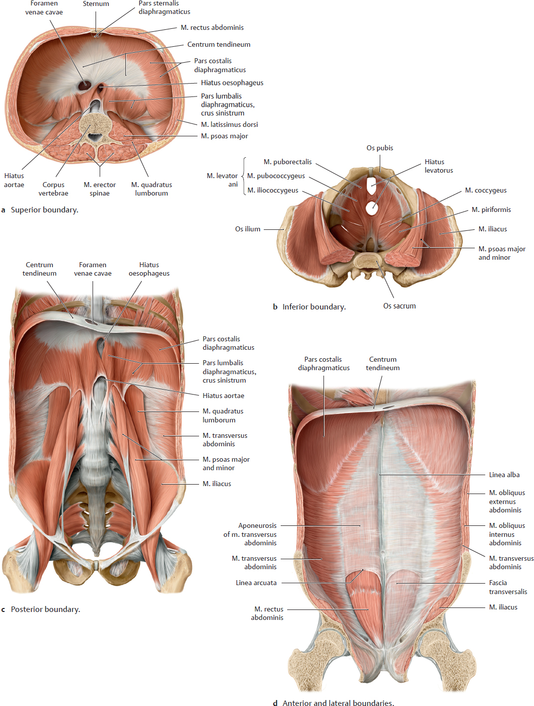
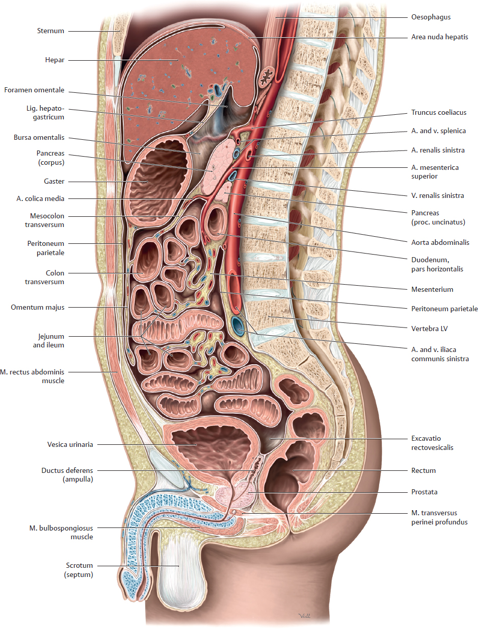
16 Structure of the Cavitas Abdominis and Cavitas Pelvis: Overview A Architecture and wall structure of the cavitas abdominis and cavitas pelvis Whereas the cavitas thoracica and cavitas abdominis are separated by the diaphragma, the cavitas abdominis and cavitas pelvis are continuous with each other. They are divided topographically by the linea terminalis. Thus, they form a single functional unit (see p. 3). Bones (columna vertebralis, cavea thoracis, and pelvis) as well as muscles (diaphragma, abdominal wall, and diaphragma pelvis muscles) along with their fasciae and aponeuroses form the walls of this space. It is bounded by the following structures: • Superiorly (see Ca): diaphragma with right and left domes and centrum tendineum • Inferiorly (see Cb): bony pelvis, muscles of the pelvic wall (m. iliacus, m. obturator internus, m. piriformis, and m. coccygeus), and mm. diaphragmatis pelvis (mainly the m. levator ani forming most of the diaphragma pelvis) • Posteriorly (see Cc): lumbar columna vertebralis, deep muscles of the abdominal wall (m. quadratus lumborum and m. psoas major), and mm. dorsi proprii • Anteriorly and laterally (see Cd): anterior and lateral muscles of the abdominal wall together with their aponeuroses (m. rectus abdominis and m. transversus abdominis as well as mm. obliqui internus and externus abdominis) B Functional aspects of abdominal and pelvic wall structure: Abdominal press Abdominal press is made possible by the structure of the abdominal and pelvic walls, which plays an important role in their elastic properties. “Abdominal press” describes the voluntary contraction of the diaphragma, and abdominal and pelvic muscles. When the muscles contract they reduce the volume of the cavitas abdominis thereby significantly raising the intra-abdominal pressure: pressure in the standing position is approximately 1.7 kPa (2.75 mmHg), when lying down it is approximately 0.2 kPa (1.5 mmHg), and under strain including coughing or squeezing it is 10–20 kPa (75–150 mmHg). Abdominal press is important in • Emptying of the rectum (defecation), of the bladder (micturition), and of the stomach (vomiting), • Uterine contractions during the expulsive phase of labor (“expulsive pains”), • Stabilizing the columna vertebralis (mainly the lumbar spine) and the trunk (the wall stiffens like the wall of an inflated ball), for example when lifting heavy loads, but also in the standing posture (hydrostatic effect of abdominal press). Hernias occur when the pressure load is greater than the strength of the complex myofascial network. They develop either in the anterior abdominal wall or more commonly in the groin region because the weight of the pelvic and abdominal organs puts increasing strain on the wall structures, which increases from superior to inferior. Additionally, the diaphragma pelvis muscles in particular are much less able to withstand the increased abdominal pressure than the abdominal wall muscles or the diaphragma. During abdominal press, closure of the glottis and the retaining of air in the lungs gives support to the diaphragma; there is no such compensatory mechanism in the diaphragma pelvis muscles making it a characteristic weak spot. After excessive stretching (e.g., caused by vaginal delivery) the diaphragma pelvis is unable to maintain the pelvic organs in their normal position (pelvic floor descent) and provides inadequate support for the abdominal press. The results are urinary and fecal incontinence. C Boundaries of the cavitas abdominis and cavitas pelvis a Diaphragma, inferior view; b Diaphragma pelvis, superior view; c (d) Anterior (posterior) view of the posterior (anterior) trunk wall. A Midsagittal section through the abdomen and pelvis, viewed from the left side
16.1 Architecture, Wall Structure, and Functional Aspects
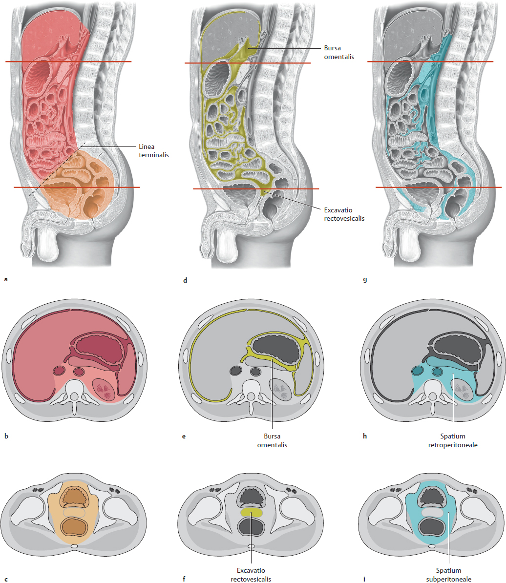
16.2 Divisions of the Cavitas Abdominis and Cavitas Pelvis
Basicmedical Key
Fastest Basicmedical Insight Engine