• Cutaneous, gastrointestinal, neuroophthalmic, musculoskeletal, renal, hematological manifestations also common
• Nonspecific elevations in transaminases, alkaline phosphatase
• Plasma vitamin A levels may be normal
Microscopic
• Stellate cell hyperplasia and hypertrophy
• Hepatocellular injury, inflammation minor
• Fibrosis begins in perisinusoidal pattern, may progress to cirrhosis
Top Differential Diagnoses
• Methotrexate therapy
• Steroid use
• IV fat emulsion administration
• Stellate cell activation has been reported in chronic viral hepatitis following transplantation
• May be marker of early fibrogenesis following transplantation
Hypervitaminosis 
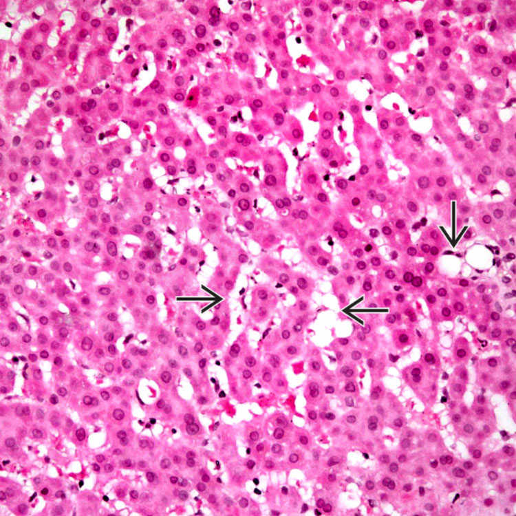
Low-power view of stellate cell hyperplasia in hypervitaminosis A illustrates bubbly, “multivacuolated” hepatic stellate cells in the sinusoids . Note the lack of inflammation or hepatocyte degeneration.
Chronic Overingestion of Vitamin A 
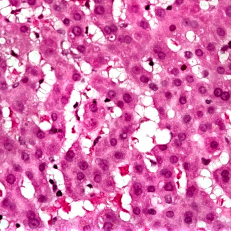
Hyperplastic, hypertrophic stellate cells are seen in the sinusoids of a patient who chronically overingested vitamin A supplements.
Hyperplastic and Hypertrophic Stellate Cells in Sinusoids 
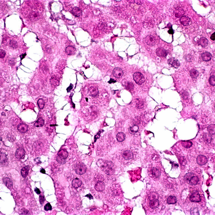
High magnification shows hyperplastic, hypertrophic stellate cells in the sinusoids with swollen, clear cytoplasm and delicate cytoplasmic processes.
Bubbly Cytoplasm of Stellate Cells This patient does not have increased perisinusoidal fibrosis, but the contrast of the trichrome stain accentuates the bubbly cytoplasm of the stellate cells.
TERMINOLOGY
Abbreviations
• Hepatic stellate cells (HSC)
Synonyms
• Ito cell
• Perisinusoidal lipocyte
Definitions
• Phenotypic changes in stellate cells as result of activation
Most commonly seen in vitamin A toxicity (hypervitaminosis A)
– Even moderate amounts of vitamin A can cause liver disease if taken over long period of time
• Stellate cells reside in space of Disse
Long cytoplasmic processes surround sinusoids
Contain small lipid droplets that are rich in vitamin A
Only gold members can continue reading. Log In or Register to continue