Glandular component may contain conventional ductal-type, clear cell, or signet ring cell components
Top Differential Diagnoses
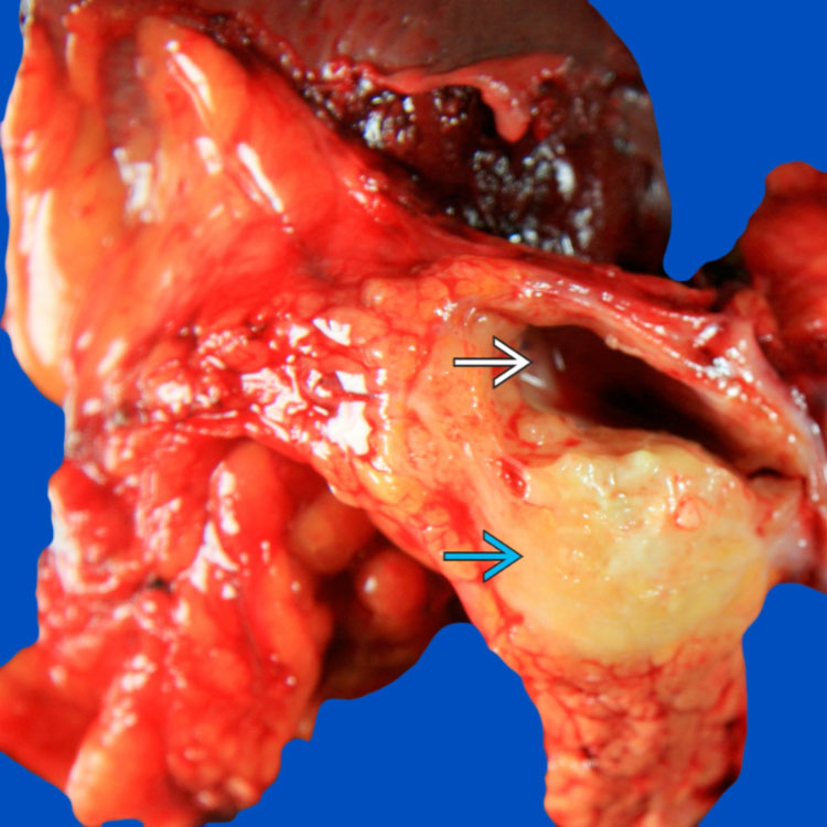
Primary adenosquamous cell carcinoma in the tail of the pancreas shows a relatively well-demarcated, tan-white to yellow solid mass
 with a cystic component
  .
.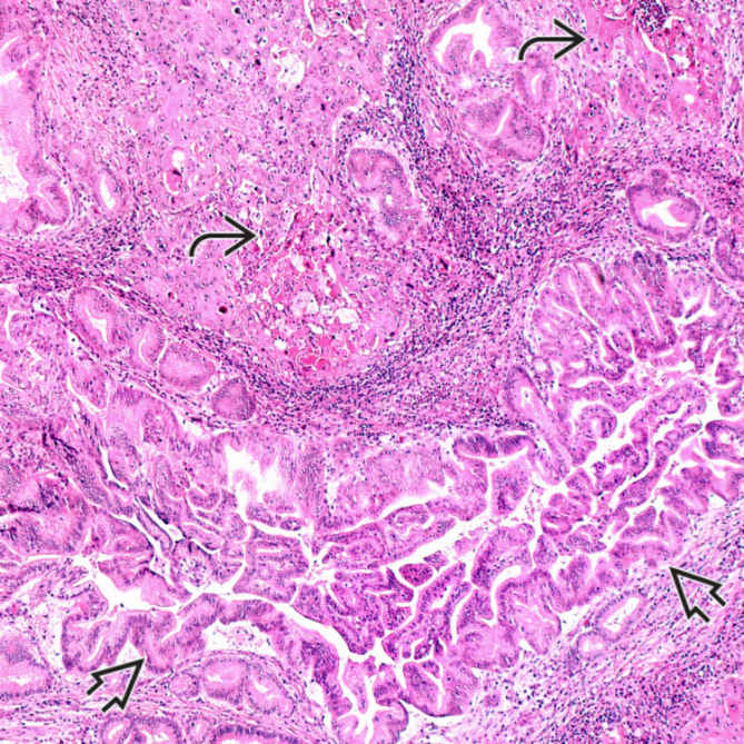
Adenosquamous cell carcinoma of the pancreas demonstrates islands of malignant squamous cells with keratinization
 adjacent to high-grade pancreatic intraepithelial neoplasia involving an interlobular duct
  .
.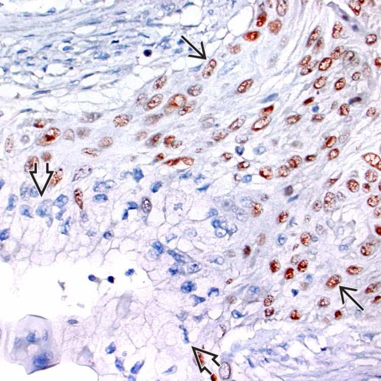
Immunohistochemical stain for p63 shows nuclear staining in the squamous component
 of adenosquamous cell carcinoma, while the glandular component is negative
  .
.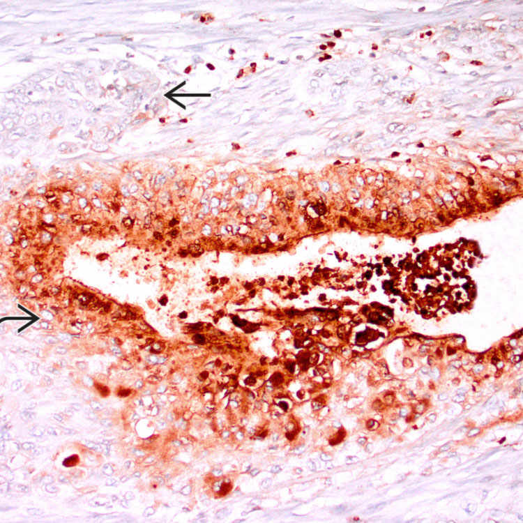
 , while the squamous component
  is negative.
ETIOLOGY/PATHOGENESIS
Neoplastic
• A few hypotheses attempt to explain origin of squamous component


Stay updated, free articles. Join our Telegram channel
Full access? Get Clinical Tree
