•
Most cases are related to UV radiation
•
Previous radiation therapy implicated in some cases; usually associated with more aggressive SCC
•
Often arises in sun-damaged skin of elderly patients (usually head and neck)
Vast majority of cases associated with preexisting actinic keratosis (AK)
•
Complete surgical excision is optimal and definitive therapy
•
Prognosis usually good in superficial and well-differentiated cases
•
Worse prognosis with poorly differentiated, deeply invasive, or aggressive subtypes
•
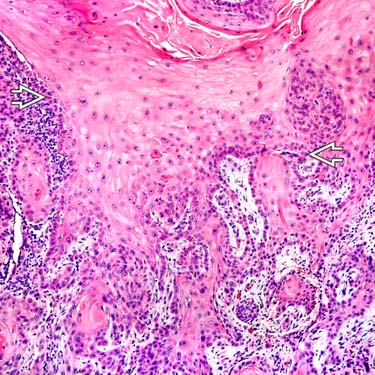
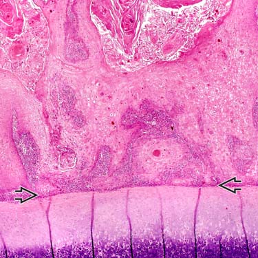
Proliferation of invasive atypical keratinocytes, often with areas of keratinization (keratin pearls) and squamous eddies
•
Cells are present in nests, sheets, and cords
•
Cytologically, cells show abundant eosinophilic cytoplasm and large nucleus with vesicular chromatin and prominent nucleoli
•
Degree of differentiation is variable, ranging from well to moderately to poorly differentiated
•
Multiple variants of differing malignant potential described
•
Poorly differentiated carcinoma (Including metastatic)
•
Pseudoepitheliomatous hyperplasia
(Courtesy S. Yashar, MD.)
•
Squamous cell carcinoma (SCC)
•
Sarcomatoid carcinoma (spindle cell carcinoma/carcinosarcoma/metaplastic carcinoma)
•
Acantholytic (adenoid/pseudoglandular) SCC
•
Verrucous carcinoma: Well-differentiated variant
•
Keratoacanthoma (KA): Very well-differentiated variant, regresses spontaneously
•
Malignant tumor of squamous keratinocytes
•
Most cases are related to UV radiation
•
Some cases are likely related to chronic inflammation (i.e., SCC arising in burns, lupus, lichen planus)
•
Previous radiation therapy is implicated in some cases; usually associated with more aggressive SCC
•
Chronic wounds and burn scars also can be associated with high-risk SCC
•
Human papillomavirus is associated with some cases
Especially verrucous carcinoma (low grade) and SCC in immunosuppressed patients (high grade)
•
Age
Usually in elderly, especially solar-related lesions
However, can present in wide age range (34-95 years)
–
Rare cases in children (should prompt genetic studies)
•
Sex
Slightly more common in males, overall
•
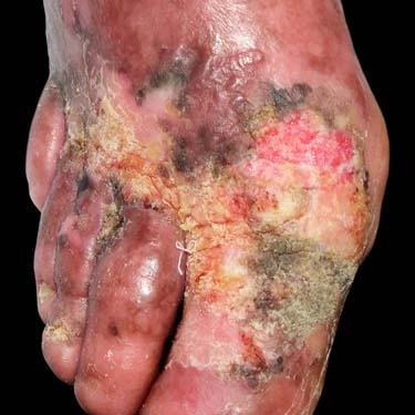
Slow-growing papular, nodular, or plaque lesion
•
Often arises in sun-damaged skin (head and neck tumors)
Vast majority of cases associated with preexisting actinic keratosis (AK)
•
May be ulcerated or bleeding
•
Ear canal and middle ear tumors may present with pain, hearing loss, and discharge
•
Surgical approaches
Complete surgical excision is optimal and definitive therapy
–
Mohs surgery has been shown to be highly effective for most tumors
•
Drugs
If patients are not surgical candidates, topical chemotherapeutics or immunomodulators may be used
•
Radiation
May be used for very advanced cases where surgical therapy is not curative
•
Usually excellent in most cases
•
Worse prognosis with poorly differentiated, deeply invasive, or rare aggressive subtypes
•
Site of tumor important for prognosis
Lip and ear tumors more aggressive, regardless of degree of differentiation
•
Papular to nodular or plaque-like lesion; can be exophytic
May be ulcerated or hemorrhagic
•
Variable; can be small or large lesions

 .
.
 (but not into) the auricular cartilage is shown.
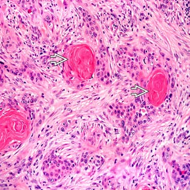
 and a sclerotic stroma with scattered inflammatory cells.


