and John E. Skandalakis1
(1)
Centers for Surgical Anatomy and Technique, Emory University School of Medicine Piedmont Hospital, Atlanta, GA, USA
Abstract
Studies indicate that the majority of the population has two splenic segments—superior and inferior. However, three and more segments have been reported in the literature. Splenic vasculature is segmental and highly variable. The splenic veins follow the pattern of the arterial distribution more consistently in the smaller branches than in the larger ones.
Splenectomy is usually performed for hemorrhage, hypersplenism, Hodgkin’s disease staging, or a problem such as an abscess, cyst, or tumor. Detailed step-by-step procedure is presented for splenectomy, ligation of the splenic pedicle, and occlusion of the splenic artery. An expanded section on laparoscopic splenectomy highlights the “leaning spleen” technique. Patients with fractures of the left 9–11th ribs should be observed closely for splenic rupture. A healthy ruptured spleen can be delivered easily from the abdomen; a large, diseased spleen requires careful dissection.
Anatomy
General Description of the Spleen
The spleen is concealed at the left hypochondrium and is not palpable under normal conditions. It is associated with the posterior portions of the left 9th, 10th, and 11th ribs—separated from them by the diaphragm and the costodiaphragmatic recess (Fig. 15.1).
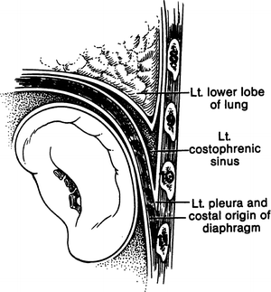

Figure 15.1.
Location of the spleen. Reproduced from JE Skandalakis, GL Colborn, LB Pemberton, et al. Prob Gen Surg 7(1):1–17, 1990. With permission from Wolters Kluwer Health.
If one divides the spleen into three parts, the upper third is related to the lower lobe of the left lung, the middle third to the left costodiaphragmatic recess, and the lower third to the left pleura and costal origin of the diaphragm.
For all practical purposes, the spleen has two surfaces: parietal and visceral (Fig. 15.2). The convex parietal surface is related to the diaphragm, and the concave visceral surface is related to the surfaces of the stomach, kidney, colon, and tail of the pancreas. On the concave hilar surface, the entrance and exit of the splenic vessels at the splenic portals in most specimens form the letter S, which is evident if one connects the upper polar, hilar, and lower polar vessels.
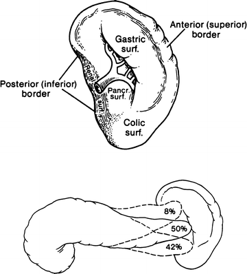

Figure 15.2.
Splenic borders. (Top) Anterior and posterior border. (Bottom) Relations of the tail of the pancreas to the spleen. Reproduced from JE Skandalakis, GL Colborn, LB Pemberton, et al. Prob Gen Surg 7(1):1–17, 1990. With permission from Wolters Kluwer Health.
A double layer of peritoneum covers the entire spleen, except for the hilum (Fig. 15.3).
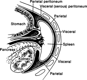

Figure 15.3.
Sagittal view of peritoneum covering the spleen. Reproduced from JE Skandalakis, GL Colborn, LB Pemberton, et al. Prob Gen Surg 7(1):1–17, 1990. With permission from Wolters Kluwer Health.
Chief Splenic Ligaments
At the hilum, the visceral peritoneum joins the right layer of the greater omentum and forms the gastrosplenic and splenorenal ligaments, the two chief ligaments of the spleen (Figs. 15.4 and 15.5). These two ligaments form the splenic pedicle. The splenic capsule is formed by the visceral peritoneum; it is as friable as the spleen itself and as easily injured (Fig. 15.4).
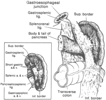
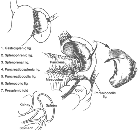

Figure 15.4.
The peritoneal attachments of the spleen. (Inset) The hilum of the spleen showing the short gastric and gastroepiploic vessels in the gastrosplenic ligament (By permission of JE Skandalakis, SW Gray, and JR Rowe. Anatomical Complications in General Surgery. New York: McGraw-Hill, 1983).

Figure 15.5.
Chief and minor splenic ligaments. Reproduced from JE Skandalakis, GL Colborn, LB Pemberton, et al. Prob Gen Surg 7(1):1–17, 1990. With permission from Wolters Kluwer Health.
The superior pole of the spleen lies close to the stomach and may be fixed to it. The inferior pole lies 5–7 cm from the stomach. The gastrosplenic ligament contains the short gastric arteries above and the left gastroepiploic vessels below; it should be incised only between clamps or preferably after the vessels are ligated one by one. Transfixion sutures may be used.
The splenorenal ligament envelops the splenic vessels and the tail of the pancreas. The outer layer of the splenorenal ligament forms the posterior layer of the gastrosplenic ligament. Careless division of the former may injure the short gastric vessels. Bleeding from these vessels may be the result of too-enthusiastic deep posterior excavation by the index and middle fingers of an operator seeking to mobilize and retract the spleen to the right. The splenorenal ligament itself is nearly avascular and may be incised, but the fingers should stop at the pedicle (Fig. 15.6).
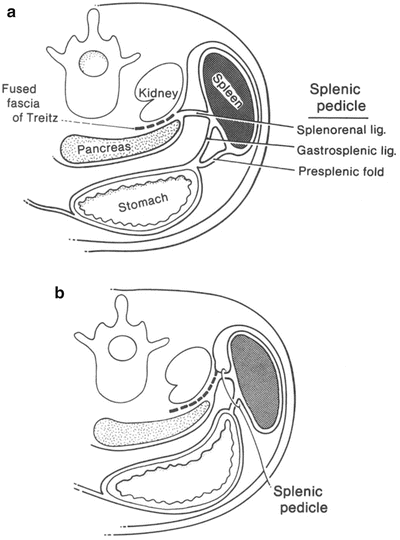

Figure 15.6.
The splenic pedicle. (a) Long pedicle with a presplenic fold. (b) Short pedicle (By permission of JE Skandalakis, SW Gray, and JR Rowe. Anatomical Complications in General Surgery. New York: McGraw-Hill, 1983).
Minor Splenic Ligaments
The spleen has several minor ligaments, and their names indicate their connections (Fig. 15.5): the splenophrenic, splenocolic, pancreaticosplenic, pancreaticocolic, and phrenicocolic ligaments and the presplenic fold. The splenophrenic ligament (Fig. 15.5) is the reflection of the leaves of the mesentery to the posterior body wall and to the inferior surface of the diaphragm at the area of the upper pole of the spleen close to the stomach. It is usually avascular, but it should be inspected for possible bleeding after section.
Tortuous or aberrant inferior polar vessels of the spleen or a left gastroepiploic artery can lie close enough to the splenocolic ligament to be injured by careless incision of the ligament, possibly resulting in massive bleeding. The ligament should be incised between clamps.
The pancreaticosplenic ligament (Fig. 15.5) is said to be present when the tail of the pancreas does not touch the spleen.
The pancreaticocolic ligament (Fig. 15.5) is the upper extension of the transverse mesocolon and is somewhat of a bridge from the tail or body of the pancreas to the splenic flexure of the colon (Fig. 15.7).
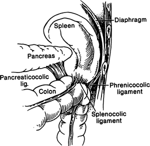

Figure 15.7.
Relation of the pancreaticocolic, phrenicocolic, and splenocolic ligaments to the transverse mesocolon. Reproduced from JE Skandalakis, GL Colborn, LB Pemberton, et al. Prob Gen Surg 7(1):1–17, 1990. With permission from Wolters Kluwer Health
The “phrenicocolic ligament” is not a splenic ligament; i.e., it is not connected to the spleen, but the spleen rests upon it. It extends between the splenic flexure of the colon and the diaphragm and constitutes the “splenic floor” (Fig. 15.5).
The presplenic fold is a peritoneal fold anterior to the gastrosplenic ligament (Fig. 15.5), often containing the left gastroepiploic vessels. Excessive traction on this fold during upper abdominal operations can result in a tear in the splenic capsule.
Vascular System of the Spleen
Splenic Artery and Its Branches
The splenic artery, in most people, is a branch of the celiac trunk, arising together with the common hepatic and left gastric arteries. The splenic artery varies in length from 8 to 32 cm and in diameter from 0.5 to 1.2 cm. The normal course of the splenic artery crosses the left side of the aorta, passes along the upper border of the pancreas reaching the tail in front, and then crosses the upper pole of the left kidney.
The left gastroepiploic artery arises most often from the splenic trunk. Less often it arises from the inferior terminal splenic branch or its branches and rarely from the middle splenic trunk or the superior terminal branch.
There is no question that the spleen can tolerate ligation of the splenic artery because of the available collateral circulation. Therefore, the spleen can be saved if necessary. Surgeons should remember that ligation of the splenic artery near its origin can result in hyperamylasemia resulting from deterioration of the pancreatic blood supply. Preoperative splenic arterial embolization as an adjunct to high-risk splenectomy has been advised.
Splenic Vein and Its Branches
The splenic vein travels with the splenic artery (Fig. 15.8), sometimes crossing over or under it. The anatomy of the splenic vein is summarized in Fig. 15.9. The patterns are highly variable, and as in the arteries, no one vein resembles the next. The single characteristic of most of the short gastric veins is that they communicate directly with the spleen, entering at its upper part, rather than through the extrasplenic venous vessels. The left gastroepiploic venous drainage is into the splenic veins.
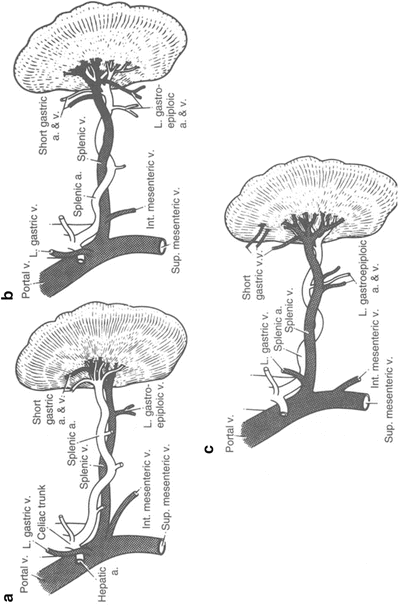
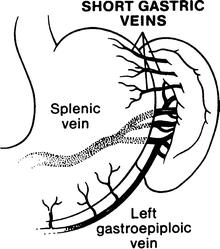

Figure 15.8.
Relation of splenic artery and splenic vein. (a) Artery anterior to vein (usual pattern). (b) Artery both anterior and posterior to vein. (c) Artery posterior to vein (least common configuration) (By permission of JE Skandalakis, SW Gray, and JR Rowe. Anatomical Complications in General Surgery. New York: McGraw-Hill, 1983)

Figure 15.9.
Anatomy of the splenic vein. Reproduced from JE Skandalakis, GL Colborn, LB Pemberton, et al. Prob Gen Surg 7(1):1–17, 1990. With permission from Wolters Kluwer Health.
Lymphatic Drainage
One of the peculiarities of the spleen is the lack of provision for lymphatics for the splenic pulp. The splenic lymphatic chain (Fig. 15.10) is reported to be formed by suprapancreatic nodes, infrapancreatic nodes, and afferent and efferent lymph vessels.
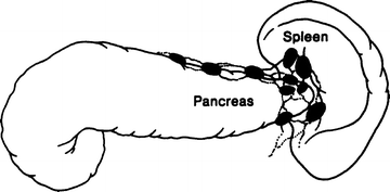

Figure 15.10.
Lymphatic drainage of the spleen. Reproduced from JE Skandalakis, GL Colborn, LB Pemberton, et al. Prob Gen Surg 7(1):1–17, 1990. With permission from Wolters Kluwer Health.
Segmental Anatomy
We propose that the term segments be used when referring to splenic parts separated by avascular planes, elsewhere called lobes. Studies indicate that the majority of the population has two splenic segments—superior and inferior. However, three and more segments have been reported in the literature.
Accessory Spleens
The reported incidence of accessory spleens in autopsies varies from approximately 20 to 30 %. In approximately 60 % of affected patients, only one accessory spleen is present. In 20 %, two such spleens are present, and in 17 %, there are three or more splenic structures. Two-thirds to three-fourths of accessory spleens are located at or near the hilum of the normal organ, and about 20 % are embedded in the tail of the pancreas. The remainder are distributed along the splenic artery, in the omentum, in the mesentery, or beneath the peritoneum (Fig. 15.11). They range from 0.2 to 10 cm in size, resembling lymph nodes or miniature spleens.
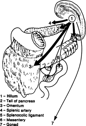

Figure 15.11.


Sites of accessory spleens in order of frequency: (1) near the splenic hilum; (2) tail of the pancreas (these contain 86–95 % of all accessory spleens); (3) omentum; (4) along the splenic artery; (5) splenocolic ligament; (6) mesentery; and (7) testis or ovary (3–7 are unusual locations) (By permission of JE Skandalakis, SW Gray, and JR Rowe. Anatomical Complications in General Surgery. New York: McGraw-Hill, 1983).
Stay updated, free articles. Join our Telegram channel

Full access? Get Clinical Tree


