S. mansoni and S. japonicum most frequently cause hepatosplenic disease


Diagnostic Checklist
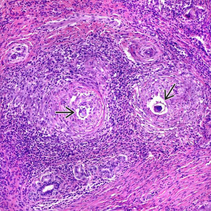
This expanded and markedly fibrotic portal area contains numerous granulomas with central ova
 ; some are clearly embryonated. There is associated chronic inflammation, but eosinophils are not prominent in this case.
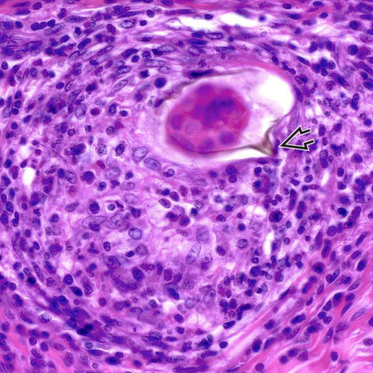
 .
.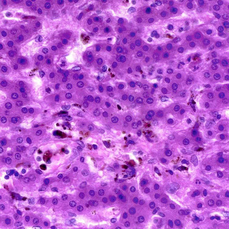
Macrophages in the portal tracts and sinusoids contain dark brown pigment, consistent with hematin, which is regurgitated by the flukes after metabolizing hemoglobin.
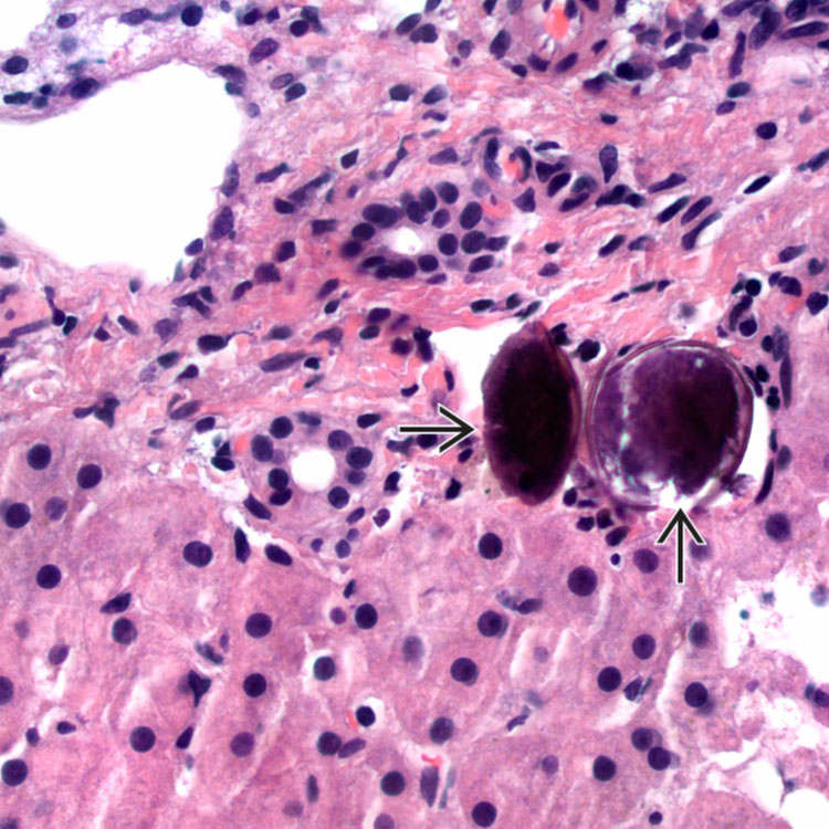
 , possibly in a small venule; the spines are not visible. In this case, a granulomatous reaction is not present. The lack of inflammation and calcification of the ova implies remote infection.
ETIOLOGY/PATHOGENESIS
Life Cycle and Infection
• Infected humans/animals contaminate fresh water with eggs by urine or feces


• Infection occurs when cercariae (infectious larvae) exit snail and penetrate skin of vertebrate host (in contaminated water)
Stay updated, free articles. Join our Telegram channel
Full access? Get Clinical Tree
